
- Title
-
Zebrafish Models of Photoreceptor Dysfunction and Degeneration
- Authors
- Noel, N.C.L., MacDonald, I.M., Allison, W.T.
- Source
- Full text @ Biomolecules
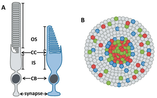
Anatomy of rod and cone photoreceptors and their organization in the human retina. (A) Cartoons of a rod (grey) and cone (blue) photoreceptor. Photoreceptors have an outer segment (OS) that is packed with light-sensitive opsin proteins, a connecting cilium (CC) that connects the OS with the mitochondria-rich inner segment (IS), a cell body (CB), and a synapse. (B) Cartoon of the human photoreceptor mosaic. Humans have three types of cones: red, green, and blue, depicted in those respective colours. The peripheral retina is rod-dense with cones interspersed throughout, while the central retina is cone-dense. |

Zebrafish photoreceptor organization. (A) Cartoon depiction of the adult zebrafish photoreceptor mosaic. UV and blue cones, depicted in purple and blue respectively, alternate in their rows while red and green double cones alternate in neighbouring rows. Rods are studded throughout. (B) Fluorescent image of a flat-mounted adult transgenic zebrafish retina, with GFP expressed in UV cones (magenta) and mCherry expressed in blue cones (cyan). The alternation of UV and blue cones in their rows is apparent. (C) Immunofluorescent image of a cryosectioned adult Tg(sws1:GFP) zebrafish retina with GFP in UV cones (green). UV opsin (red) and rhodopsin (magenta) are labelled, as well as nuclei (cyan). |