
Figure 1
- ID
- ZDB-FIG-210128-122
- Publication
- Noel et al., 2021 - Zebrafish Models of Photoreceptor Dysfunction and Degeneration
- Other Figures
- All Figure Page
- Back to All Figure Page
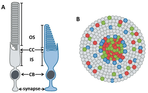
Anatomy of rod and cone photoreceptors and their organization in the human retina. (A) Cartoons of a rod (grey) and cone (blue) photoreceptor. Photoreceptors have an outer segment (OS) that is packed with light-sensitive opsin proteins, a connecting cilium (CC) that connects the OS with the mitochondria-rich inner segment (IS), a cell body (CB), and a synapse. (B) Cartoon of the human photoreceptor mosaic. Humans have three types of cones: red, green, and blue, depicted in those respective colours. The peripheral retina is rod-dense with cones interspersed throughout, while the central retina is cone-dense. |