
|
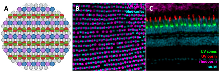
Figure 2 Zebrafish photoreceptor organization. (A) Cartoon depiction of the adult zebrafish photoreceptor mosaic. UV and blue cones, depicted in purple and blue respectively, alternate in their rows while red and green double cones alternate in neighbouring rows. Rods are studded throughout. (B) Fluorescent image of a flat-mounted adult transgenic zebrafish retina, with GFP expressed in UV cones (magenta) and mCherry expressed in blue cones (cyan). The alternation of UV and blue cones in their rows is apparent. (C) Immunofluorescent image of a cryosectioned adult Tg(sws1:GFP) zebrafish retina with GFP in UV cones (green). UV opsin (red) and rhodopsin (magenta) are labelled, as well as nuclei (cyan).