
- Title
-
lncRNA NR2F1-AS1 promotes breast cancer angiogenesis through activating IGF-1/IGF-1R/ERK pathway
- Authors
- Zhang, Q., Li, T., Wang, Z., Kuang, X., Shao, N., Lin, Y.
- Source
- Full text @ J. Cell. Mol. Med.
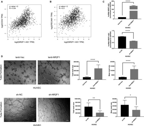
High lncRNA NR2F1‐AS1 expression is associated with breast cancer angiogenesis. A, Pearson's correlation analysis of lncRNA NR2F1‐AS1 and CD34 in breast cancer. B, Pearson's correlation analysis of lncRNA NR2F1‐AS1 and CD31 in breast cancer. C, Expression of lncRNA NR2F1‐AS1 in HUVECs overexpressing lncRNA NR2F1‐AS1 and knock‐down of lncRNA NR2F1‐AS1. D, Tube formation of transfected HUVECs (representative image). * |

lncRNA NR2F1‐AS1 promotes breast cancer angiogenesis in vitro. A, Expression of lncRNA NR2F1‐AS1 in MDA‐MB‐231 cells knock‐down of lncRNA NR2F1‐AS1 and MCF‐7 cells overexpressing lncRNA NR2F1‐AS1. B, Proliferation of HUVECs at 48 h in the TCM from transfected MDA‐MB‐231 cells and at 96 h in the TCM from transfected MCF‐7 cells. C, Tube formation of HUVECs at 6 h in the TCM from transfected MDA‐MB‐231 and MCF‐7 cells (representative image). D, Wound healing of HUVECs at 48 h in the TCM from transfected MDA‐MB‐231 and MCF‐7 cells (representative image). * |
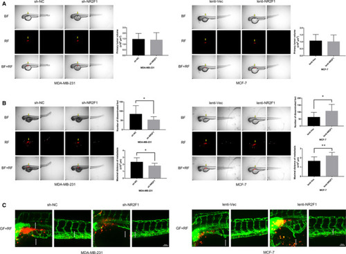
lncRNA NR2F1‐AS1 promotes breast cancer angiogenesis in zebrafish model. A, 0 h after microinjection of transfected MDA‐MB‐231 and MCF‐7 cells into the perivitelline space of embryos at 48 h post‐fertilization (representative image). B, Metastasis of the transfected breast cancer cells in zebrafish at 24 h post‐injection (representative image, for MDA‐MB‐231, n = 13/group, for MCF‐7, n = 14/group). C, Representative image of neo‐vascularization around breast cancer cells in zebrafish at 24 h post‐injection. Yellow arrows direct to the primary location of breast cancer cells. Small arrowheads direct to disseminated and metastatic tumour foci. White arrows direct to new formed tumour vascularization. BF, bright field; GF, green fluorescence; RF, red fluorescence. * |
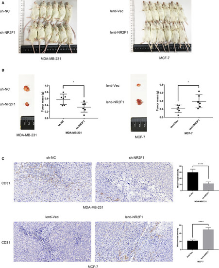
lncRNA NR2F1‐AS1 promotes breast cancer angiogenesis in mouse model. A, 28 d after implantation of transfected MDA‐MB‐231 and MCF‐7 cells in NOD/SCID mice (n = 6/group). B, Tumour size and weight at 28 d after implantation in mice (representative image). C, CD31 staining and MVD in tumour mass at 28 d after implantation in mice (representative image). * |
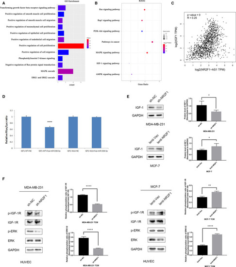
lncRNA NR2F1‐AS1 promotes breast cancer angiogenesis via IGF‐1/IGF‐1R/ERK pathway. A, GO enrichment of the target genes of miRNA‐338‐3p. B, KEGG analysis of the target genes of miRNA‐338‐3p. C, Pearson's correlation analysis of lncRNA NR2F1‐AS1 and IGF‐1. D, Dual‐luciferase reporter gene assay of miRNA‐338‐3p and IGF‐1. E, Protein expression of IGF‐1 in transfected MDA‐MB‐231 and MCF‐7 cells (representative image) and the Western blot analysis. F, Phosphorylation of IGF‐1R and ERK1/2 in HUVECs cultivated in TCM from transfected breast cancer cells (representative image) and the Western blot analysis. * |
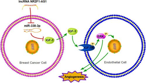
Schematic diagram of lncRNA NR2F1‐AS1 promoting breast cancer angiogenesis through IGF‐1/IGF‐1R/ERK pathway via sponging miRNA‐338‐3p |