FIGURE SUMMARY
Title

Paclitaxel-induced peripheral neuropathy is caused by epidermal ROS and mitochondrial damage through conserved MMP-13 activation

Authors
Cirrincione, A.M., Pellegrini, A.D., Dominy, J.R., Benjamin, M.E., Utkina-Sosunova, I., Lotti, F., Jergova, S., Sagen, J., Rieger, S.
Source
Full text @ Sci. Rep.

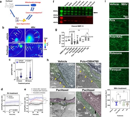
Mitochondrial ROS contribute to MMP-13 expression and axon degeneration. (a) Is mitochondrial damage involved in paclitaxel-induced axon degeneration? (b) Ratiometric images showing HyPer oxidation (arrows) in the caudal fin of larval zebrafish (dashed lines) after 3 and 48 hr of treatment (2 and 4dpf, respectively) with either 0.09%DMSO vehicle or 23 µM paclitaxel. Keratinocytes are mosaically labeled following transient injection of tp63:HyPer into 1-cell stage embryos. High oxidation is represented in red and low oxidation in blue (n = 2–3 biological replicates with 5–7 fish per group). (c) Quantification reveals increased oxidation after short and long-term treatment with paclitaxel, n = 10–15 animals. (d,e) 30–60 min quantifications of HyPer oxidation in epidermal cells expressing krt4:HyPer following treatment for 5 (d) and 48 hr (e), n = 3–6 animals in each treatment group, SEM. Paclitaxel treatment significantly elevates H2O2 levels. Comparison with H2O2 induced by fin amputation shows continuous versus transient H2O2 elevation. (f,g) Western blot (black arrow indicates cleaved MMP-13, 48 kDa) (f) and quantitative analysis (g) shows increased MMP-13 expression following treatment with paclitaxel and the mitochondrial ROS inducer paraquat but not rotenone. Antioxidants (NAC and DPI) reduce MMP-13 levels induced by paclitaxel treatment (replicate number is indicated by dots in the graph, each replicate contained pools of 10 fish). (h) TEM of (6dpf) zebrafish keratinocytes following 96 hr treatment with either 0.09% DMSO vehicle, 23 µM paclitaxel + DB04760 (MMP-13 inhibitor) or paclitaxel alone shows an intact basement membrane (yellow arrowheads) in vehicle and paclitaxel + DB04760 treated fish, whereas the basement membrane is discontinuous (white arrowheads) and large gaps (black arrows) appear where unmyelinated sensory axons normally reside (n = 3 animals per treatment group). (i) Epidermal unmyelinated sensory axons in the distal tail fin following 96 hr treatment with vehicle, paclitaxel, paclitaxel + NAC, rotenone, or paraquat. Quantification of treatments shows paclitaxel, rotenone, and paraquat induce axon degeneration whereas NAC co-administration prevents degeneration, n = 2 biological replicates, 5–7 animals per treatment group, SEM. Abbreviations: NAC = N-acetylcysteine, Pctx = paclitaxel, Rot = rotenone, PQ = paraquat, B = basal keratinocyte, BM = basement membrane, Ac = actinotrichia, N = nucleus, P = periderm, M = mitochondrion, dpf = days post fertilization.

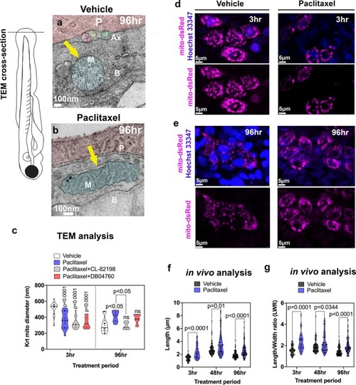
Keratinocyte mitochondria are damaged by paclitaxel treatment. (a) TEM analysis of zebrafish larvae treated for 96 hr with either 0.09% DMSO vehicle or 23 µM paclitaxel (2–6dpf). A mitochondrion (M) is shown with outer and inner membrane (white arrow) and clearly visible cristae following vehicle treatment. Three axons (Ax) are embedded between the periderm and basal keratinocyte layer. (b) The outer membrane of a mitochondrion following paclitaxel treatment is less apparent, and the space between inner and outer membrane has increased (yellow arrow). The cristae are less electron-dense, and large dark puncta are visible. (c) Quantification shows decreased mitochondria diameters upon 3 hr paclitaxel treatment with and without MMP-13 inhibitors, CL-82198 or D04760, compared with controls. 96 hr treatment shows increased mitochondria diameters in paclitaxel but not MMP-13 inhibitor treated animals (n = 3 animals per treatment group). (d,e) In vivo imaging of mitochondria in keratinocytes labeled with mito-dsRed and nuclei labeled with live Hoechst 33347 stain following treatment for 3 hr and 96 hr with either vehicle (d) or paclitaxel (e). Short and long-term treatment with paclitaxel promotes mitochondria filamentation. (f,g) Quantification after 3 hr, 48 hr and 96 hr treatment shows increased length (f) and length/width ratios (g) with paclitaxel compared to controls, n = 5–8 animals in 3 biological replicates. Abbreviations: B = basal keratinocyte, P = periderm, M = mitochondrion, Ax = axon.

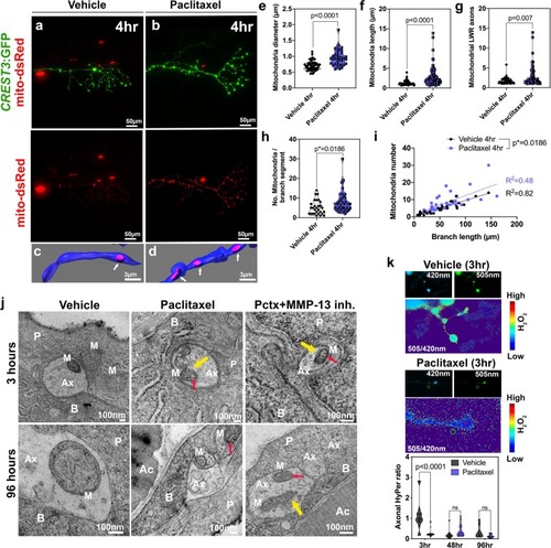
Axonal mitochondria are vacuolized following paclitaxel treatment but ROS/H2O2 levels are not elevated. (a,b) DsRed-labeled mitochondria in unmyelinated axons of somatosensory neurons labeled with CREST3:GFP following 4 hr treatment with vehicle (a) or paclitaxel (b). (c,d) Respective 3D reconstructions of axon branch segment (blue) in the epidermis containing mitochondria (pink, arrows). (ei) Quantification of axonal mitochondria shows increased diameter (e), length (f), length-width ratio (g) and number of mitochondria per branch segment (h,i), n = 5 animals per treatment group. (j) TEM analysis of axonal mitochondria (M) following treatment with 0.09% DMSO vehicle, paclitaxel with or without MMP-13 inhibitor (Cl-82198 top and DB04760 bottom) for 3 hr and 96 hr shows rapid paclitaxel-induced mitochondria vacuolization (red arrows) and membrane disruptions (yellow arrows), persisting up to 96 hr, similar to paclitaxel + MMP-13 inhibitor. (k) Axonal HyPer oxidation is elevated after DMSO treatment for 3 hr but not after prolonged treatment with either DMSO or paclitaxel, n = 5–7 animals per treatment group. Abbreviations: B = basal keratinocyte, P = periderm, M = mitochondrion, Ax = axon, Ac = Actinotrichia.

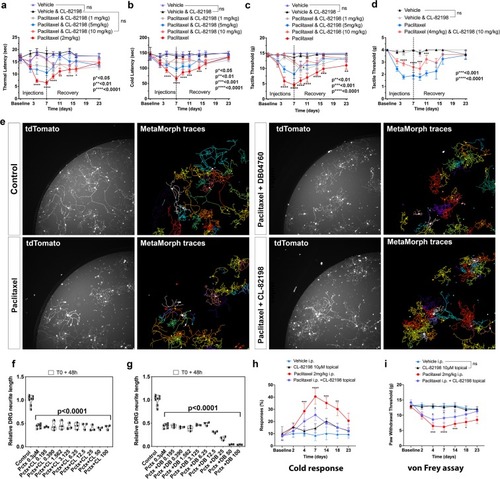
MMP-13 prevents paclitaxel-induced PIPN in rodents by neuron-extrinsic mechanism. (ad) Increased sensitivity to heat (a), cold (b) and touch (c,d) following paclitaxel treatment of rats (ac) and mice (d) compared with controls and upon injection of paclitaxel + CL-82198, n = 5–6 animals per treatment group and 5 replicates. (eg) Fields of live imaged tdTomato + DRG neurons and neurite tracing (e) and length quantification (f,g) show decreased length upon treatment with either paclitaxel, paclitaxel (0.3 µM) + CL-82198 (10 µM), or paclitaxel (0.3 µM) + DB04760 (10 µM). Data represent mean and SEM from 3 independent experiments. (h,i) Increased cold response and tactile sensitivity is alleviated upon topical application of CL-82198 to the hind paw of paclitaxel-injected rats (n = 5 animals per treatment group).

Acknowledgments
This image is the copyrighted work of the attributed author or publisher, and ZFIN has permission only to display this image to its users. Additional permissions should be obtained from the applicable author or publisher of the image. Full text @ Sci. Rep.