IMAGE

Figure 3

ID
ZDB-IMAGE-200314-15
Source
Figures for Cirrincione et al., 2020
Image
Figure Caption

Figure 3

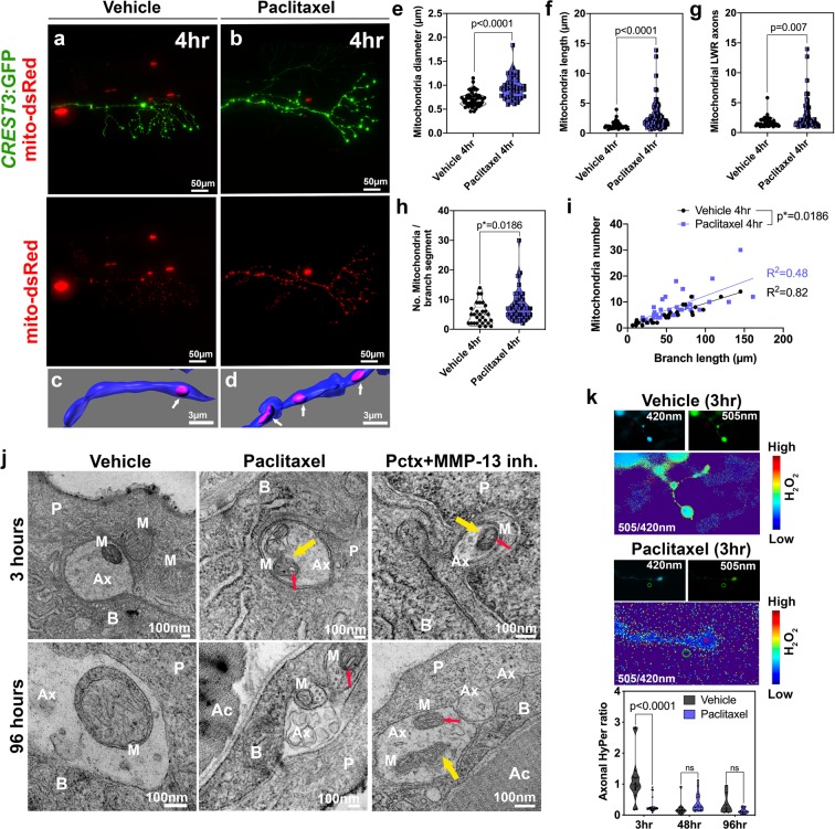
Axonal mitochondria are vacuolized following paclitaxel treatment but ROS/H2O2 levels are not elevated. (a,b) DsRed-labeled mitochondria in unmyelinated axons of somatosensory neurons labeled with CREST3:GFP following 4 hr treatment with vehicle (a) or paclitaxel (b). (c,d) Respective 3D reconstructions of axon branch segment (blue) in the epidermis containing mitochondria (pink, arrows). (ei) Quantification of axonal mitochondria shows increased diameter (e), length (f), length-width ratio (g) and number of mitochondria per branch segment (h,i), n = 5 animals per treatment group. (j) TEM analysis of axonal mitochondria (M) following treatment with 0.09% DMSO vehicle, paclitaxel with or without MMP-13 inhibitor (Cl-82198 top and DB04760 bottom) for 3 hr and 96 hr shows rapid paclitaxel-induced mitochondria vacuolization (red arrows) and membrane disruptions (yellow arrows), persisting up to 96 hr, similar to paclitaxel + MMP-13 inhibitor. (k) Axonal HyPer oxidation is elevated after DMSO treatment for 3 hr but not after prolonged treatment with either DMSO or paclitaxel, n = 5–7 animals per treatment group. Abbreviations: B = basal keratinocyte, P = periderm, M = mitochondrion, Ax = axon, Ac = Actinotrichia.

Acknowledgments
This image is the copyrighted work of the attributed author or publisher, and ZFIN has permission only to display this image to its users. Additional permissions should be obtained from the applicable author or publisher of the image. Full text @ Sci. Rep.