IMAGE

Figure 1

ID
ZDB-IMAGE-200314-13
Source
Figures for Cirrincione et al., 2020
Image
Figure Caption

Figure 1

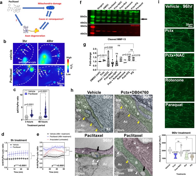
Mitochondrial ROS contribute to MMP-13 expression and axon degeneration. (a) Is mitochondrial damage involved in paclitaxel-induced axon degeneration? (b) Ratiometric images showing HyPer oxidation (arrows) in the caudal fin of larval zebrafish (dashed lines) after 3 and 48 hr of treatment (2 and 4dpf, respectively) with either 0.09%DMSO vehicle or 23 µM paclitaxel. Keratinocytes are mosaically labeled following transient injection of tp63:HyPer into 1-cell stage embryos. High oxidation is represented in red and low oxidation in blue (n = 2–3 biological replicates with 5–7 fish per group). (c) Quantification reveals increased oxidation after short and long-term treatment with paclitaxel, n = 10–15 animals. (d,e) 30–60 min quantifications of HyPer oxidation in epidermal cells expressing krt4:HyPer following treatment for 5 (d) and 48 hr (e), n = 3–6 animals in each treatment group, SEM. Paclitaxel treatment significantly elevates H2O2 levels. Comparison with H2O2 induced by fin amputation shows continuous versus transient H2O2 elevation. (f,g) Western blot (black arrow indicates cleaved MMP-13, 48 kDa) (f) and quantitative analysis (g) shows increased MMP-13 expression following treatment with paclitaxel and the mitochondrial ROS inducer paraquat but not rotenone. Antioxidants (NAC and DPI) reduce MMP-13 levels induced by paclitaxel treatment (replicate number is indicated by dots in the graph, each replicate contained pools of 10 fish). (h) TEM of (6dpf) zebrafish keratinocytes following 96 hr treatment with either 0.09% DMSO vehicle, 23 µM paclitaxel + DB04760 (MMP-13 inhibitor) or paclitaxel alone shows an intact basement membrane (yellow arrowheads) in vehicle and paclitaxel + DB04760 treated fish, whereas the basement membrane is discontinuous (white arrowheads) and large gaps (black arrows) appear where unmyelinated sensory axons normally reside (n = 3 animals per treatment group). (i) Epidermal unmyelinated sensory axons in the distal tail fin following 96 hr treatment with vehicle, paclitaxel, paclitaxel + NAC, rotenone, or paraquat. Quantification of treatments shows paclitaxel, rotenone, and paraquat induce axon degeneration whereas NAC co-administration prevents degeneration, n = 2 biological replicates, 5–7 animals per treatment group, SEM. Abbreviations: NAC = N-acetylcysteine, Pctx = paclitaxel, Rot = rotenone, PQ = paraquat, B = basal keratinocyte, BM = basement membrane, Ac = actinotrichia, N = nucleus, P = periderm, M = mitochondrion, dpf = days post fertilization.

Acknowledgments
This image is the copyrighted work of the attributed author or publisher, and ZFIN has permission only to display this image to its users. Additional permissions should be obtained from the applicable author or publisher of the image. Full text @ Sci. Rep.