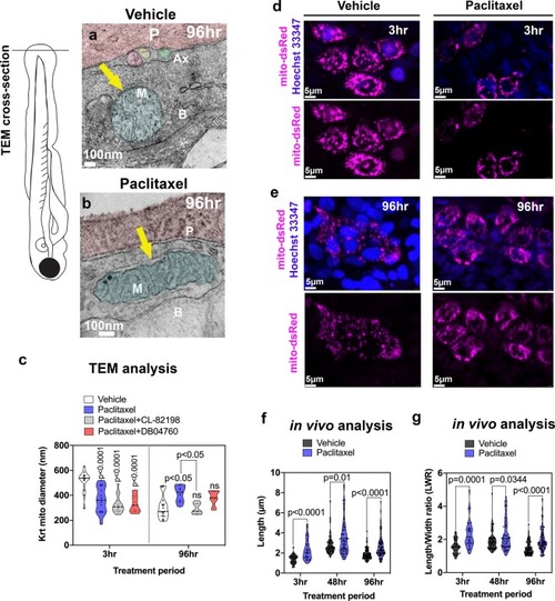
Keratinocyte mitochondria are damaged by paclitaxel treatment. (a) TEM analysis of zebrafish larvae treated for 96 hr with either 0.09% DMSO vehicle or 23 µM paclitaxel (2–6dpf). A mitochondrion (M) is shown with outer and inner membrane (white arrow) and clearly visible cristae following vehicle treatment. Three axons (Ax) are embedded between the periderm and basal keratinocyte layer. (b) The outer membrane of a mitochondrion following paclitaxel treatment is less apparent, and the space between inner and outer membrane has increased (yellow arrow). The cristae are less electron-dense, and large dark puncta are visible. (c) Quantification shows decreased mitochondria diameters upon 3 hr paclitaxel treatment with and without MMP-13 inhibitors, CL-82198 or D04760, compared with controls. 96 hr treatment shows increased mitochondria diameters in paclitaxel but not MMP-13 inhibitor treated animals (n = 3 animals per treatment group). (d,e) In vivo imaging of mitochondria in keratinocytes labeled with mito-dsRed and nuclei labeled with live Hoechst 33347 stain following treatment for 3 hr and 96 hr with either vehicle (d) or paclitaxel (e). Short and long-term treatment with paclitaxel promotes mitochondria filamentation. (f,g) Quantification after 3 hr, 48 hr and 96 hr treatment shows increased length (f) and length/width ratios (g) with paclitaxel compared to controls, n = 5–8 animals in 3 biological replicates. Abbreviations: B = basal keratinocyte, P = periderm, M = mitochondrion, Ax = axon.
|
