
- Title
-
Pectoral Fin Anomalies in tbx5a Knockdown Zebrafish Embryos Related to the Cascade Effect of N-Cadherin and Extracellular Matrix Formation
- Authors
- Lu, J.K., Tsai, T.C., Lee, H., Hsia, K., Lin, C.H., Lu, J.H.
- Source
- Full text @ J Dev Biol
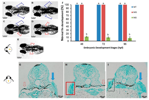
Phenotypes of pectoral fins in tbx5a-deficient embryos. Wild-type embryos with bilateral normal development of the pectoral fin (A,B). Unilateral hypoplasia (C), unilateral agenesis (D), and bilateral agenesis (E) of the pectoral fin in tbx5a morphants at 96 hpf. The rate of normal fin development was significantly reduced in tbx5a-deficient embryos at 48, 72, and 96 hpf (F). a, b: A significant difference was detected by one-way ANOVA with Duncan’s multiple range test. WT: Uninjected group, MIS: Control MO-injected group, MO: tbx5a morphant group. (G) Cross section of embryos with morphologic unilateral agenesis of the pectoral fin with Alcian blue staining showing well-organized glycosaminoglycans (GAGs) in the normal pectoral fin (blue arrow) with complete absence of chondrocytes and GAGs at the aplasia site (red arrow). (H) Cross section of the embryo with morphologic bilateral hypoplasia/agenesis of the pectoral fin showing the absence of chondrocytes and GAGs at the aplasia site (red arrow) with disorganized GAGs in the hypoplastic pectoral fin (red box). (I) Cross section of the embryo with morphologic unilateral hypoplasia of the pectoral fin showing disorganized GAGs in the hypoplastic pectoral fin (black box) and well-organized GAGs in the normal pectoral fin (blue arrow).
|
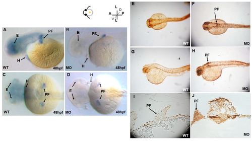
Lateral (A) and dorsal (C) views of tbx5a gene expression in the eyes, heart, and bilateral pectoral fins of wild-type embryos at 48 hpf. tbx5a gene expression in the eyes, heart, and pectoral fins was severely inhibited at 48 hpf in both the lateral (B) and ventral (D) views. Detection of apoptotic cells by the TUNEL assay in wild-type (E,G,I) and tbx5a knockdown (F,H,J) zebrafish embryos at 37 hpf. No TUNEL-positive cells are observable in the dorsal view (E), lateral view (G), or cross sections at the pectoral fin level (I) in wild-type embryos. However, massive numbers of TUNEL-positive cells in the pectoral fin region (black arrow) in the tbx5a morphant embryos were visible in the dorsal view (F), lateral view (H) and cross section at the pectoral fin level (J). WT, Uninjected group; MO, tbx5a morphant group; PF, pectoral fin; E, eye, H, heart.
|
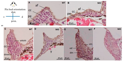
Cross section of a pectoral fin stained with HE. Wild-type embryos with well-developed myocytes and organized chondrocytes in pectoral fins at 37 (A), 48 (C), and 72 hpf (E). The hypoplastic pectoral fin of the tbx5a morphant showed reduced myocytes and disorganized chondrocytes at 37 (B), 48 (D) and 72 hpf (F). (af, apical fold; cc, chondrogenic condensation; dm, dorsal musculature; dmm, dorsal myogenic mesenchyme; ed. endoskeleton disc; vm, ventral musculature; vmm, ventral myogenic mesenchyme; pe, peritoneal epithelium; y, yolk, WT, Uninjected group, MO, tbx5 morphant group).
|
Cross section of pectoral fins stained with Alcian blue. Wild-type embryos showed well-developed apical folds and organized chondrocytes at 37 (A), 48 (C), and 72 hpf (E). The tbx5a morphant showed severe pectoral fin hypoplasia with loss of the apical fold, reduced chondrocytes, unorganized chondrogenic condensation and endoskeletal disc at 37 (B), 48 (D), and 72 hpf (F). (af, apical fold; cc, chondrogenic condensation; dm, dorsal musculature; dmm, dorsal myogenic mesenchyme; ed. endoskeleton disc; vm, ventral musculature; vmm, ventral myogenic mesenchyme; pe, peritoneal epithelium; y, yolk).
|
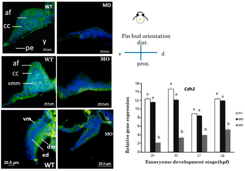
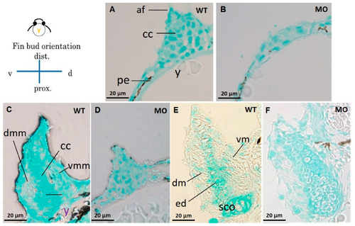
Cross sections of pectoral fins subjected to immunofluorescence staining for CDH2. Pectoral fins of wild-type embryos showed abundant N-cadherin surrounding chondrocytes at 37 (A) and 48 hpf (C). N-cadherin is observable in the dorsal and ventral muscular region surrounding the endoskeletal disc at 72 hpf (E). The tbx5amorphant with a stubby pectoral fin showed a lack of N-cadherin at 37 hpf (B), 48 hpf (D) and 72 hpf (F). cdh2 expression was significantly inhibited in the tbx5 morphant group at 26, 30, 37, and 48 hpf (G) (n = 50 embryos, triplet). (cl, cleithrum; cc, chondrogenic condensation; dm, dorsal musculature; ed, endoskeleton disc; sco, scapulocoracoid; vm, ventral musculature; blue, nuclear; green, CDH2 protein, WT: Uninjected group, MIS: Control MO-injected group, MO: tbx5 morphant group, a, b: A significant difference was detected by one-way ANOVA with Duncan’s multiple range test.
|

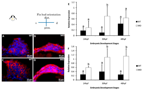
The immunochemical characterization of fibronectin and HA-binding protein in fin tissue at 48 hpf. Compared with wild-type embryos (A,C), tbx5a MO embryos showed decreased expression of fibronectin and HA-binding protein at 48 hpf (B,D). Cx43 expression in the tbx5a morphant group was significantly inhibited at 24, 30, 36, and 48 hpf (n = 50, triplet) (E). a, b: A significant difference was detected by One-way ANOVA with Duncan’s multiple range test; red in (A,B), fibronectin; green in (C,D), HA-binding protein; blue: nuclear, WT: Uninjected group, MIS: Control MO-injected group, MO: tbx5a morphant group.
|
Immunohistochemical staining of apoptosis-related proteins. Pectoral fins were stained with apoptosis-related (BAD and BCL2) antibodies (red) and counterstained with DAPI (blue) to observe the nucleus. BAD (B) and BCL2 (D) proteins were significantly uninhibited in the tbx5a morphant group compared with those in the Uninjected group (A,C). The embryo anterior is shown on the left. The Bad (E) and Bcl2 (F) expression in the tbx5a morphant group was significantly uninhibited at 24, 30, 36, and 48 hpf (n = 50, triplet) (E). hpf, hours postfertilization; WT, Uninjected group; MO, tbx5a morphant group. a, b: A significant difference was detected by a, b: A significant difference was detected by One-way ANOVA with Duncan’s multiple range test.
|