
Image
|
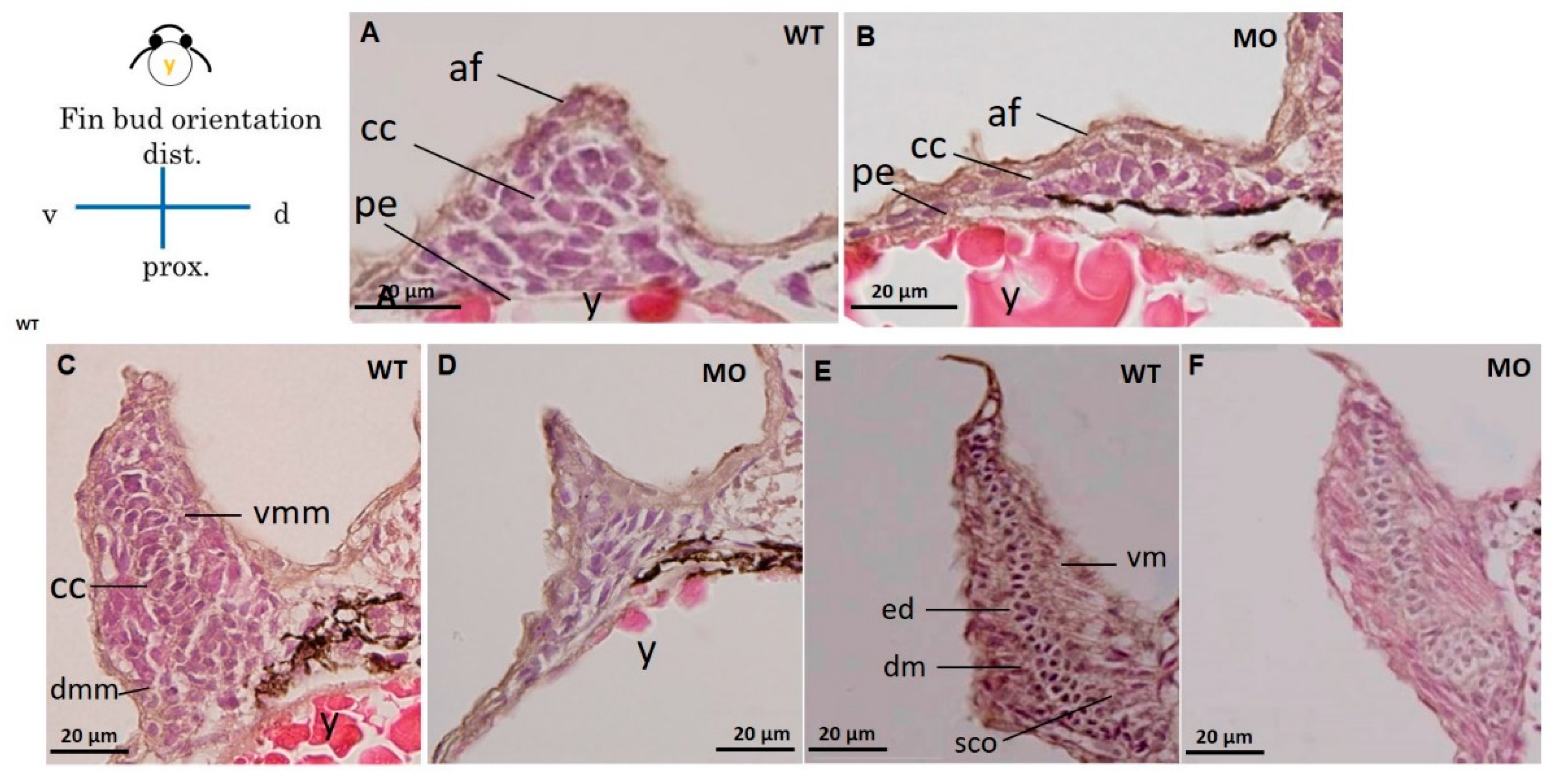
Figure Caption
Fig. 3
Cross section of a pectoral fin stained with HE. Wild-type embryos with well-developed myocytes and organized chondrocytes in pectoral fins at 37 (A), 48 (C), and 72 hpf (E). The hypoplastic pectoral fin of the tbx5a morphant showed reduced myocytes and disorganized chondrocytes at 37 (B), 48 (D) and 72 hpf (F). (af, apical fold; cc, chondrogenic condensation; dm, dorsal musculature; dmm, dorsal myogenic mesenchyme; ed. endoskeleton disc; vm, ventral musculature; vmm, ventral myogenic mesenchyme; pe, peritoneal epithelium; y, yolk, WT, Uninjected group, MO, tbx5 morphant group).
Acknowledgments
This image is the copyrighted work of the attributed author or publisher, and
ZFIN has permission only to display this image to its users.
Additional permissions should be obtained from the applicable author or publisher of the image.
Full text @ J Dev Biol