
|
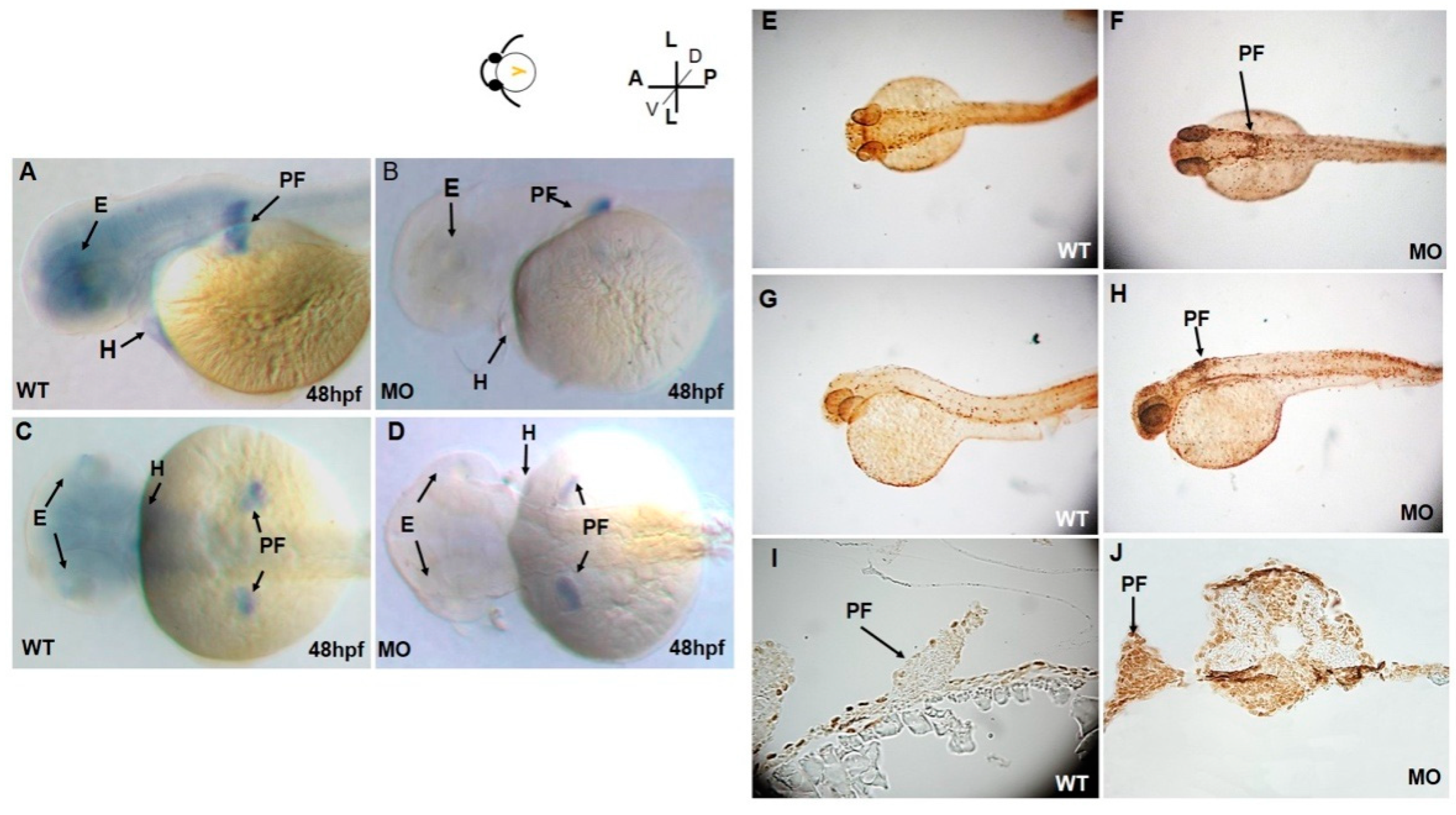
Fig. 2
Lateral (A) and dorsal (C) views of tbx5a gene expression in the eyes, heart, and bilateral pectoral fins of wild-type embryos at 48 hpf. tbx5a gene expression in the eyes, heart, and pectoral fins was severely inhibited at 48 hpf in both the lateral (B) and ventral (D) views. Detection of apoptotic cells by the TUNEL assay in wild-type (E,G,I) and tbx5a knockdown (F,H,J) zebrafish embryos at 37 hpf. No TUNEL-positive cells are observable in the dorsal view (E), lateral view (G), or cross sections at the pectoral fin level (I) in wild-type embryos. However, massive numbers of TUNEL-positive cells in the pectoral fin region (black arrow) in the tbx5a morphant embryos were visible in the dorsal view (F), lateral view (H) and cross section at the pectoral fin level (J). WT, Uninjected group; MO, tbx5a morphant group; PF, pectoral fin; E, eye, H, heart.