FIGURE SUMMARY
Title

Inhibition of let-7c Regulates Cardiac Regeneration after Cryoinjury in Adult Zebrafish

Authors
Narumanchi, S., Kalervo, K., Perttunen, S., Wang, H., Immonen, K., Kosonen, R., Laine, M., Ruskoaho, H., Tikkanen, I., Lakkisto, P., Paavola, J.
Source
Full text @ J Cardiovasc Dev Dis

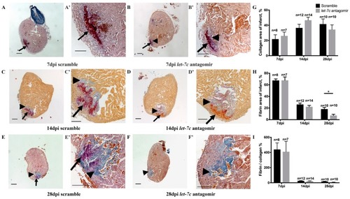
Collagen and fibrin quantified with AFOG staining at 7 dpi, 14 dpi and 28 dpi. Intact myocardium is stained in orange, fibrin in red and collagen in blue (AF); higher magnifications of the panels on the left (A’F’). Arrows point to fibrin and arrowheads to collagen (AF; A’F’). Collagen (G), fibrin (H) and fibrin/collagen% (I) (n = 7 for 7 dpi antagomir, n = 8 for 7 dpi scramble, n = 14 for 14 dpi antagomir, n = 12 for 14 dpi scramble, n = 10 for 28 dpi antagomir and scramble). Scale bar: 200 μm. * p ≤ 0.05.

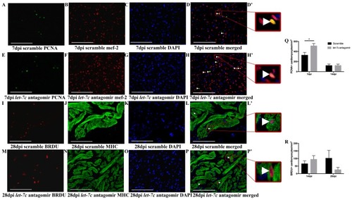
Quantification of proliferating cardiomyocytes in cryoinjured hearts. Representative images at 7 dpi of PCNA, mef-2, DAPI, merged image and inset of merged image in scramble (AD,D’) and in let-7c antagomir hearts (EH,H’). Representative images at 28 dpi of BRDU, MHC, DAPI, merged image and inset of merged image in scramble (IL,L’) and in let-7c antagomir hearts (MP,P’). Positive cardiomyocytes marked with arrowheads. In PCNA images (A–H’) mef-2 marks cardiomyocytes in red, PCNA positive cells marked in green and DAPI nuclei marked in blue. In BRDU images (IP’), MHC marks myocardium in green, BRDU positive cells marked in red and DAPI nuclei marked in blue. Insets are magnified images of merged image (D’,H’,L’,P’). Newly formed cardiomyocytes are quantified by PCNA staining at 7 dpi and 14 dpi (Q) and by BRDU labeling at 14 dpi and 28 dpi (R). (PCNA: n = 3 for 7 dpi antagomir and scramble, n = 8 for 14 dpi antagomir and scramble. BRDU: n = 10 for 14 dpi antagomir, n = 9 for 14 dpi scramble, n = 8 for 28 dpi antagomir and n = 6 for 28 dpi scramble). Scale bar: 100 μm. * p ≤ 0.05.

Echocardiography to assess cardiac function. The epicardial border is marked (A). FVS measured from recorded B-mode videos before cryoinjury, and at 1 dpi, 7 dpi, 14 dpi, 21 dpi, and 28 dpi (B). (n = 60 for before, n = 18 for 1 dpi, n = 29 for 7 dpi antagomir, n = 30 for 7 dpi scramble, n = 28 for 14 dpi antagomir, n = 29 for 14 dpi scramble, n = 16 for 21 dpi antagomir, n = 18 for 21 dpi scramble, n = 16 for 28 dpi antagomir and n = 17 for 28 dpi scramble). * p ≤ 0.05, *** p ≤ 0.0005.

Acknowledgments
This image is the copyrighted work of the attributed author or publisher, and ZFIN has permission only to display this image to its users. Additional permissions should be obtained from the applicable author or publisher of the image. Full text @ J Cardiovasc Dev Dis