
- Title
-
Resveratrol Ameliorates Glucocorticoid-Induced Bone Damage in a Zebrafish Model
- Authors
- Luo, Q., Liu, S., Xie, L., Yu, Y., Zhou, L., Feng, Y., Cai, .
- Source
- Full text @ Front Pharmacol

Influence of dexamethasone (Dex) concentration on zebrafish skull. Schematic diagram of TG(SP7:EGFP) zebrafish larvae (A,B). IOD values of green fluorescence of Dex-induced bone damage in TG zebrafish larvae (C). Images of green fluorescence in TG zebrafish larvae skull [in profile (D)]. The Dex concentrations used were 0, 0.25, 5.00, 10.00, 15.00, 20.00, and 25.00 μmol/l, and 0.1% DMSO was used as the control. n = 15, ***p < 0.01. PHENOTYPE:
|
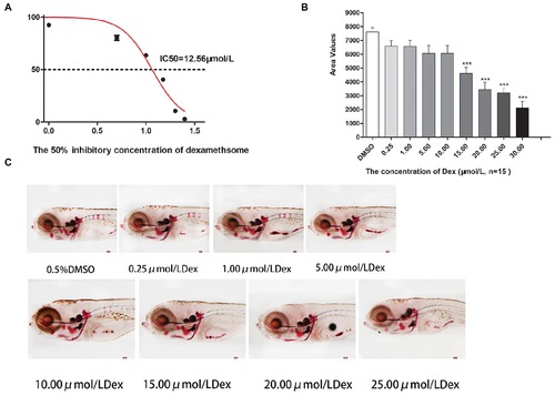
IC50 and area of bone mineralization after Dex-induced bone damage in AB-strain zebrafish larvae. The 50% inhibitory concentration of dexamethasone (Dex) (A). Analysis of mineralization area in the skull and spine of Dex-treated AB-strain zebrafish larvae at 9 dpf (B). Alizarin red S staining of the skull in Dex-treated AB-strain zebrafish larvae at 9 dpf (C). The Dex concentrations used were 0.25, 1.00, 5.00, 10.00, 15.00, 20.00, and 25.00 μmol/l. The zebrafish larvae were exposed to Dex from 3 to 9 dpf, and 0.1% DMSO served as the control. n = 15, *p < 0.05, ***p < 0.01. PHENOTYPE:
|
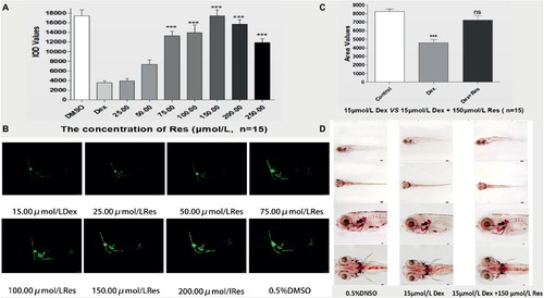
Effect of Res on Dex-induced bone damage in zebrafish. IOD values of green fluorescence of Res after Dex-induced bone damage in TG zebrafish larvae (A). Images of green fluorescence in TG zebrafish larvae skull [in profile (B)]. The Res concentrations used were 25.00, 50.00, 75.00, 100.00, 150.00, 200.00, and 250.00 μmol/l, and 0.1% DMSO served as the control. The area of bone mineralization after Res treatment of Dex-induced bone damage in AB-strain zebrafish larvae at 9 dpf [(C) 15.00 μmol/l Dex vs. 15.00 μmol/l Dex plus 150.00 μmol/l Res]. Alizarin red S staining of the skull after Res treatment of Dex-induced bone damage in AB-strain zebrafish larvae at 9 dpf (D). n = 15, *p < 0.05, ***p < 0.01. PHENOTYPE:
|

ZFIN is incorporating published figure images and captions as part of an ongoing project. Figures from some publications have not yet been curated, or are not available for display because of copyright restrictions. PHENOTYPE:
|