- Title
-
Nonpathological Chondrogenic Features of Valve Interstitial Cells in Normal Adult Zebrafish
- Authors
- Schulz, A., Brendler, J., Blaschuk, O., Landgraf, K., Krueger, M., Ricken, A.M.
- Source
- Full text @ J. Histochem. Cytochem.
|
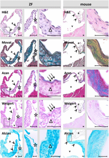
Atrioventricular (AV) valves of young adult (1-year old) zebrafish (ZF, left) and mice (right). AV valve leaflets are outlined by arrow heads. Large polygonal-shaped cells (triangles) are the main structural constituent of the base and mid-region of AV valves in ZF. In contrast, collagen-rich connective tissue without layering represents the basic structural unit in the basal aspects of AV valves in mice. In ZF, collagen and elastic material (arrows) is enriched in a thin layer on the atrial (inflow) aspect of the AV valve. An ubiquitous strong Alcian blue staining demonstrates presence of a proteoglycan/glycosaminoglycan-rich matrix. A trabecular band anchors to the ZF valve tip and extend down in the ventricle (star). Insets indicate the regions magnified on the further right photographs. Russel-Movat’s pentachrome stain (Movat), Azan stain, Weigert’s resorcin-fuchsin stain, and Alcian blue stain. Scale bar equals 25 µm in each photograph. Abbreviations: H&E, hematoxylin-eosin; a, atrium; v, ventricle. |
|
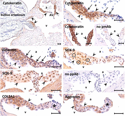
Immunohistochemical characterization of the tissue composing the base and mid-region of atrioventricular (AV) valves in young adult (1-year old) zebrafish (ZF). Heart valve leaflets are outlined by arrow heads. Immunohistochemistry reveals co-expression of epithelial (cytokeratin) and mesenchymal (vimentin) markers, SOX-9 staining in the nuclei (circles), and type 2a1 collagen and aggrecan-based matrix formation by the central layer of large polygonal-shaped cells. In contrast, the endocardial cells of the valves show vimentin expression, whereas cytokeratin is lacking (arrows). Type 2a1 collagen shows more intracellular than extracellular staining. Aggrecan antibody staining is occasionally thickened into irregular interstitial clumps. Of note, immunoreactivity in particular for type 2a1 collagen and aggrecan is almost absent in the apical tip region of the valves (asterisk). Scale bar equals 25 µm in each photograph. Abbreviations: a, atrium; v, ventricle; COL2A1, type 2a1 collagen; No pmAb, no primary monoclonal antibody controls; No ppAB, no primary polyclonal antibody controls. |
|
Immunohistochemical characterization of ZF atrioventricular (AV) valves (outlined by arrow heads) at different ages. The atrium is indicated by an “a.” At the juvenile age (35 dpf), AV valves are populated by small oval-shaped cells. At the young adult age (4 months old) a mixture of larger polygonal-shaped cells is visible. In even older AV valves (>1-year old), large polygonal-shaped cells dominate the basal and mid-region aspect of AV valves. PCNA positivity (arrows) is predominantly observed at the juvenile age along the subendothelial aspect of the AV valves. Discrete cytokeratin staining in the juvenile AV valves indicates that the VICs forming the central cells of adult valves may have retained epithelial characteristics from previous stages. SOX-9 and type 2a1 collagen staining is associated with the transition of the cells to the large polygonal shape. Squares indicate the regions magnified on the further down photographs. Scale bar equals 25 µm in each photograph. Abbreviations: ZF, zebrafish; PCNA, proliferating cell nuclear antigen; VIC, valve interstitial cell; H&E, hematoxylin-eosin; COL2A1, type 2a1 collagen. |
|
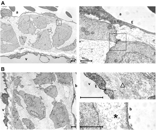
Transmission electron microscopic analysis of the basal aspects of the atrioventricular (AV) (A) and bulboventricular (BV) (B) valves in an adult (1-year old) zebrafish. The atrium is indicated by an “a,” the ventricle by a “v,” and the bulbus by a “b.” The large polygonal-shaped cells of the central cell layer are loosely arranged and only occasionally linked by cell-cell contacts (square). The cells show a central nucleus and an organelle-poor cytoplasm with few mitochondria. The extracellular matrix (ECM) around the circumference of the central cell layer is structured differently at the atrial and ventricular aspects and ventricular and bulbar aspects of AV and BV valves, respectively. At the atrial and ventricular aspect of AV and BV valves, respectively, the ECM is rich in fibril-like structures (triangle) in agreement with the strong histological staining for type 2a1 collagen. At the ventricular and bulbar aspect of AV and BV valves, respectively, it is relatively homogeneous and essentially free from visible fibril-like structures (asterisk). Scale bar equals 1 µm in each photograph. Abbreviation: E, endocardial lining. |


