Fig. 1
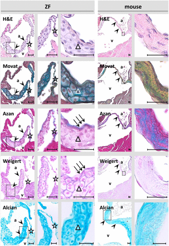
Atrioventricular (AV) valves of young adult (1-year old) zebrafish (ZF, left) and mice (right). AV valve leaflets are outlined by arrow heads. Large polygonal-shaped cells (triangles) are the main structural constituent of the base and mid-region of AV valves in ZF. In contrast, collagen-rich connective tissue without layering represents the basic structural unit in the basal aspects of AV valves in mice. In ZF, collagen and elastic material (arrows) is enriched in a thin layer on the atrial (inflow) aspect of the AV valve. An ubiquitous strong Alcian blue staining demonstrates presence of a proteoglycan/glycosaminoglycan-rich matrix. A trabecular band anchors to the ZF valve tip and extend down in the ventricle (star). Insets indicate the regions magnified on the further right photographs. Russel-Movat’s pentachrome stain (Movat), Azan stain, Weigert’s resorcin-fuchsin stain, and Alcian blue stain. Scale bar equals 25 µm in each photograph. Abbreviations: H&E, hematoxylin-eosin; a, atrium; v, ventricle.

