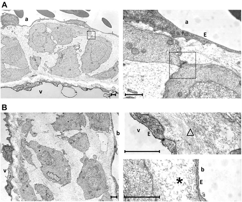
Fig. 4
Transmission electron microscopic analysis of the basal aspects of the atrioventricular (AV) (A) and bulboventricular (BV) (B) valves in an adult (1-year old) zebrafish. The atrium is indicated by an “a,” the ventricle by a “v,” and the bulbus by a “b.” The large polygonal-shaped cells of the central cell layer are loosely arranged and only occasionally linked by cell-cell contacts (square). The cells show a central nucleus and an organelle-poor cytoplasm with few mitochondria. The extracellular matrix (ECM) around the circumference of the central cell layer is structured differently at the atrial and ventricular aspects and ventricular and bulbar aspects of AV and BV valves, respectively. At the atrial and ventricular aspect of AV and BV valves, respectively, the ECM is rich in fibril-like structures (triangle) in agreement with the strong histological staining for type 2a1 collagen. At the ventricular and bulbar aspect of AV and BV valves, respectively, it is relatively homogeneous and essentially free from visible fibril-like structures (asterisk). Scale bar equals 1 µm in each photograph. Abbreviation: E, endocardial lining.

