
|
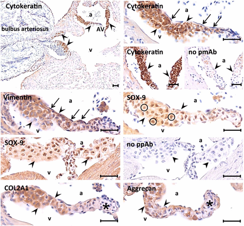
Fig. 2
Immunohistochemical characterization of the tissue composing the base and mid-region of atrioventricular (AV) valves in young adult (1-year old) zebrafish (ZF). Heart valve leaflets are outlined by arrow heads. Immunohistochemistry reveals co-expression of epithelial (cytokeratin) and mesenchymal (vimentin) markers, SOX-9 staining in the nuclei (circles), and type 2a1 collagen and aggrecan-based matrix formation by the central layer of large polygonal-shaped cells. In contrast, the endocardial cells of the valves show vimentin expression, whereas cytokeratin is lacking (arrows). Type 2a1 collagen shows more intracellular than extracellular staining. Aggrecan antibody staining is occasionally thickened into irregular interstitial clumps. Of note, immunoreactivity in particular for type 2a1 collagen and aggrecan is almost absent in the apical tip region of the valves (asterisk). Scale bar equals 25 µm in each photograph. Abbreviations: a, atrium; v, ventricle; COL2A1, type 2a1 collagen; No pmAb, no primary monoclonal antibody controls; No ppAB, no primary polyclonal antibody controls.