
- Title
-
Pannexin 1 Is Critically Involved in Feedback from Horizontal Cells to Cones
- Authors
- Cenedese, V., de Graaff, W., Csikós, T., Poovayya, M., Zoidl, G., Kamermans, M.
- Source
- Full text @ Front. Mol. Neurosci.
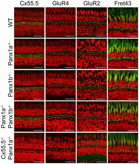
Overview of marker proteins for outer retinal function. Immunocytochemical staining for Cx55.5 (horizontal cells (HCs) gap-junctions), GluR4 (glutamate receptors on OFF-BC dendrites), GluR2 (glutamate receptors on HC dendrites) and Fret43 (synaptic terminals of double cones). Expression of these proteins did not differ between WT and the various mutant animals (Pax1a−/−, Panx1b−/−, Panx1a−/−/Panx1b−/−, Cx55.5−/−/Panx1a−/−). Scale bar is 25 μm. |

Expression of NTPDase1 in the outer retina does not differ between WT and the various genotypes (Pax1a−/−, Panx1b−/−, Panx1a−/−/Panx1b−/−, Cx55.5−/−/Panx1a−/−). Scale bar is: 25 μm. EXPRESSION / LABELING:
|

Light induced feedback responses in cones. Cones were saturated with a small spot of light and voltage clamped at −50 mV. A full field white light stimulus was applied. This resulted a small inward current: the light induced feedback response. (A) The black traces represent the mean light induced feedback response in WT. The red traces show the mean response of the various mutants. When Panx1a or Panx1b was mutated, feedback responses reduce but seem to keep a fast light-onset response. However, in the Cx55.5−/−/Panx1a−/− animals the response on-set seems to be slowed down. (B) Quantification of the sustained feedback response for all the mutants. All mutants showed a significantly reduced feedback response. *p < 0.05. PHENOTYPE:
|
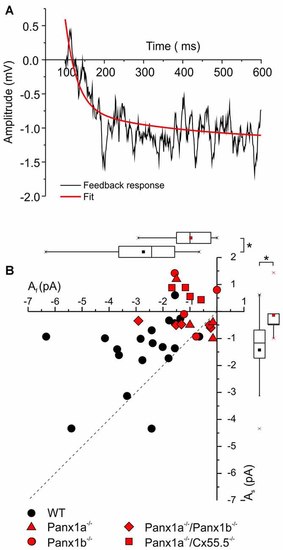
Quantification of the two feedback components. A double exponential function was fitted through the feedback responses (See “Materials and Methods” section). (A) Representative feedback trace (black) with fitted curve (red). (B) Scatter plot of the fast (x-axis) and the slow (y-axis) feedback component for WT (black symbols) and the Panx1−/− animals combined (red symbols). The dotted line indicates equal amplitude for the fast and the slow feedback component. All the WT points are above this line, indicating that the amplitude of the fast feedback component is the largest. In the Panx1−/− animals both the fast and the slow feedback component are significantly reduced. *p < 0.05. PHENOTYPE:
|
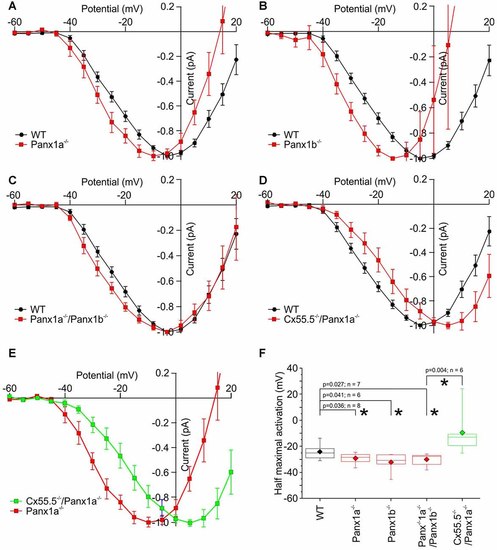
Normalized IV-relations of the cone Ca-current in WT and the various mutant animals. (A–C) In all Panx1 mutants the half activation potential of the Ca-current shifts to negative potentials. (D) In the Cx55.5−/−/Panx1a−/− animals, the Ca-current shift slightly to positive potentials. (E) The activation function of ICa has shifted strongly to positive potentials in the Cx55.5−/−/Panx1a−/− animals compared to the Panx1a−/− animals (Red curve). (F) Quantification of the shifts of the Ca-current in cones. *p < 0.05. |