
IMAGE
Fig. 2
- ID
- ZDB-IMAGE-180523-19
- Antibodies
- Publication
- Cenedese et al., 2017 - Pannexin 1 Is Critically Involved in Feedback from Horizontal Cells to Cones
- All Figures
- Figures for Cenedese et al., 2017
Image
|
Figure Caption
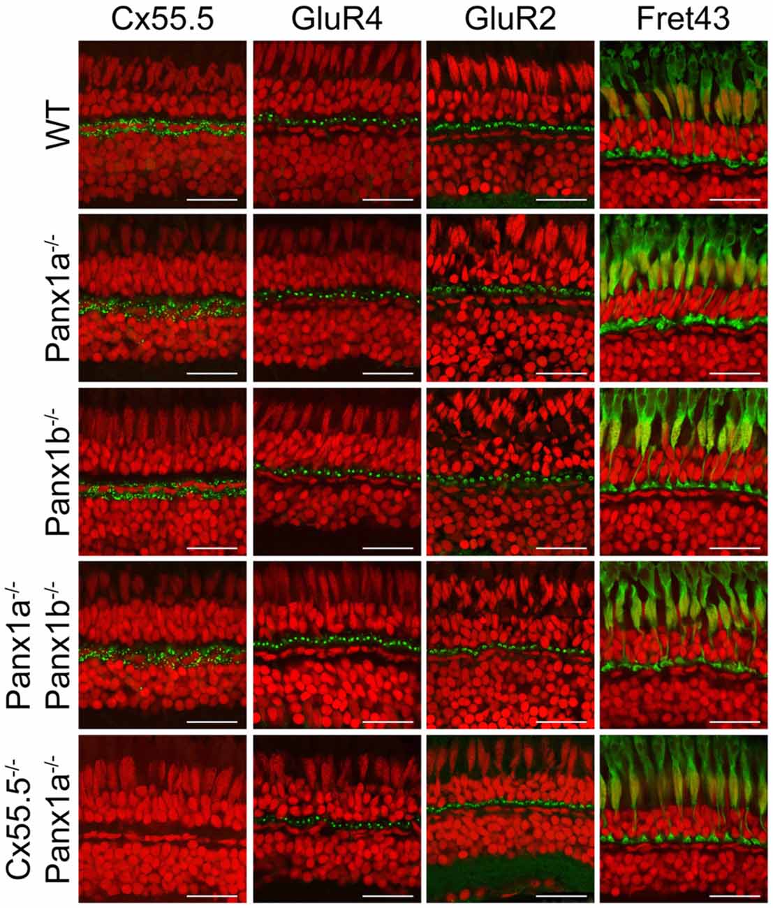
Fig. 2
Overview of marker proteins for outer retinal function. Immunocytochemical staining for Cx55.5 (horizontal cells (HCs) gap-junctions), GluR4 (glutamate receptors on OFF-BC dendrites), GluR2 (glutamate receptors on HC dendrites) and Fret43 (synaptic terminals of double cones). Expression of these proteins did not differ between WT and the various mutant animals (Pax1a−/−, Panx1b−/−, Panx1a−/−/Panx1b−/−, Cx55.5−/−/Panx1a−/−). Scale bar is 25 μm.
Figure Data
Acknowledgments
This image is the copyrighted work of the attributed author or publisher, and
ZFIN has permission only to display this image to its users.
Additional permissions should be obtained from the applicable author or publisher of the image.
Full text @ Front. Mol. Neurosci.