
- Title
-
Neurogenesis-Promoting Natural Product α-Asarone Modulates Morphological Dynamics of Activated Microglia
- Authors
- Cai, Q., Li, Y., Mao, J., Pei, G.
- Source
- Full text @ Front. Cell. Neurosci.
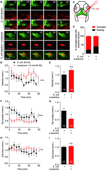
α-asarone affects microglial morphological dynamics in vivo. E. coli was inject into the gap the optic tectum region (A) of the transgenic Tg(Apo-E:eGFP) zebrafish. Time-lapse images were taken at 5-min intervals (B). Zebrafish pre-incubated with α-asarone showed decreased activation, n = 3 (C). The pre-treatment did not cause significant effect to the average migration velocity (E), but the dynamics of velocity change was significant (D). Results were normalized to “0 min” value. α-asarone treatment resulted significant declines in both dynamical changes and average numbers of tips (F,G). α-asarone treatment did not affect the cell area (H,I). (*p < 0.05, unpaired two-tailed Student t-test for independent data comparison vs. E. coli injection; ***p < 0.001, two-way ANOVA for group comparison between the dynamical trends, the numbers were on the bars). |
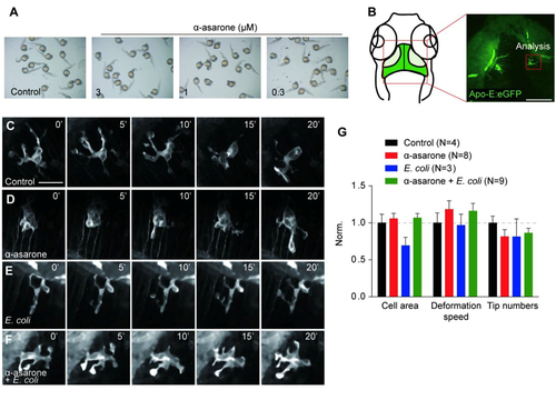
Effect of α-asarone and E. coli on the zebrafish embryonic development and resting microglial morphological dynamics The Tg(Apo-E:eGFP) transgenic zebrafish embryos were treated with 0.3-3 µM α-asarone from 12 hours post fertilisation (hpf). The developmental status was examined at 5 days post fertilisation (dpf) and revealed no defect under α-asarone treatment (0.3-3µM) (A). Real-time live imaging of microglia cells from the optic tectum region of a 5-dpf larva (B) and captured morphological dynamics of resting microglia at 5-minute intervals. (C) Untreated, (D) 3µM α-asarone, (E) E. coli injection, (F) E. coli and α-asarone (3µM) co-treatment. The results showed no significant effect of E. coli and α-asarone to the resting microglial cell area, deformation speed and tip numbers (G). |