
- Title
-
Zebrafish as a new model to study effects of periodontal pathogens on cardiovascular diseases
- Authors
- Widziolek, M., Prajsnar, T.K., Tazzyman, S., Stafford, G.P., Potempa, J., Murdoch, C.
- Source
- Full text @ Sci. Rep.

ZFIN is incorporating published figure images and captions as part of an ongoing project. Figures from some publications have not yet been curated, or are not available for display because of copyright restrictions. |
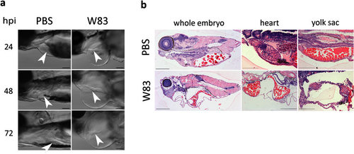
Effect of wild-type Pg W83 on zebrafish larvae tissue structure. Lateral view of impaired, elongated heart morphology of Pg W83-infected larvae; white arrowhead indicates heart. Heart chambers were distant and much smaller in comparison to PBS-injected control larvae (a). Sagittal histological sections of H&E stained 48 hpi Pg W83-infected larvae revealing advanced tissue damage in the cranial and cardiac regions along with yolk sac oedemas (b). In all figures larvae were infected with 5 × 10⁴ CFU Pg at 30 hpf W83 or PBS as a control. At least three individual experiments were performed and images are representative of at least n = 5 larvae per group in each experiment. Scale bars = 100 μm in (a) and 200 μm in (b). |
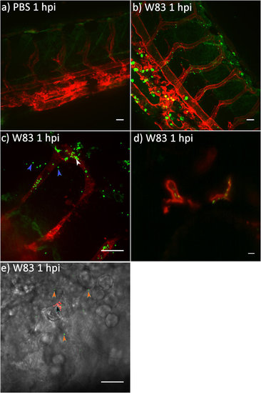
Pg W83 disseminates rapidly in zebrafish larvae and is able to cross the vascular barrier to invade surrounding tissues. Confocal images of kdrl:memRFP transgenic zebrafish larvae at 1 hpi following systemic infection with PBS control (a) or fluorescein-labelled 5 × 10⁴ CFU Pg W83 (b,c). Fluorescent Pg W83 was co-localised to the red-labelled endothelium (white arrowhead in c) and cross the vasculature into the tissue (blue arrowheads in c). Light sheet image of fluorescein-labelled Pg W83 adherent to the heat tissues of zebrafish larvae (d). Pg are rapidly phagocytosed; high-power image of phagocytosed Pg within a phagocyte (red, black arrowhead) and non-phagocytosed Pg (green, orange arrowhead) (e). Scale bar = 20 μm. Images are representative of two independent experiments showing similar findings. EXPRESSION / LABELING:
|
|
ZFIN is incorporating published figure images and captions as part of an ongoing project. Figures from some publications have not yet been curated, or are not available for display because of copyright restrictions. |
|
ZFIN is incorporating published figure images and captions as part of an ongoing project. Figures from some publications have not yet been curated, or are not available for display because of copyright restrictions. |

Gingipains are crucial virulence factors of Pg in the zebrafish larvae infection model. Kaplan-Meier survival plots of zebrafish larvae infected with Pg W83 (WT), ΔRgpA deficient mutant, ΔRgpB deficient mutant, ΔKgp deficient mutant, Kgp and RgpA deficient mutant (ΔK/Ra) or gingipain null mutant (ΔK/Ra-b) (a). Percentage of dead, oedematous or healthy WT Pg W83 or gingipain mutant-infected larvae after 24 (b) and 72 hpi (c) respectively. Morphology of zebrafish larvae 72 hpi with WT or ΔK/Ra-b mutant of Pg W83 (d). PBS was injected as a control. All experiments were performed at least 3 times. Bars represent means ± SD. In all experiments WT Pg or its gingipain mutants were injected into 30 hpf zebrafish larvae at 5 × 10⁴ CFU. Black arrow indicates oedemas in WT Pg W83-infected larvae. Scale bar = 500 μm. Comparisons between survival curves were made using the log rank test. Differences between groups displaying oedema were measured using One-way ANOVA after normality was assured. |
|
ZFIN is incorporating published figure images and captions as part of an ongoing project. Figures from some publications have not yet been curated, or are not available for display because of copyright restrictions. |

Confocal images of zebrafish larvae at 1 and 24 hpi following systemic infection with 5 x 10⁴ CFU fluorescein and pHrodo labelled Pg W83. Small green dots represent non-phagocytosed Pg whilst small red dots are phagocytosed Pg. The large red staining in the PBS controls in the tail image is due to increased pigmentation along the back of the larvae at this stage or larvae development. Similar pigmentation was observed in the eye at 24 hpi masking the presence of Pg in this tissue and so this data is not shown. Images show the presence of less Pg in the tissues at 24 hpi with increased levels of phagocytosis. W83 Pg were injected into 30 hpf zebrafish larvae at 5 x 10⁴ CFU in each experiment. Scale bar = 50μm. |