
|
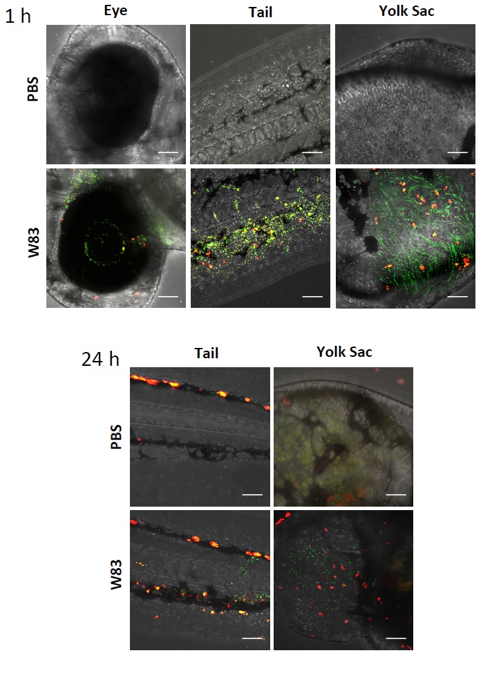
Fig. S1
Confocal images of zebrafish larvae at 1 and 24 hpi following systemic infection with 5 x 10⁴ CFU fluorescein and pHrodo labelled Pg W83. Small green dots represent non-phagocytosed Pg whilst small red dots are phagocytosed Pg. The large red staining in the PBS controls in the tail image is due to increased pigmentation along the back of the larvae at this stage or larvae development. Similar pigmentation was observed in the eye at 24 hpi masking the presence of Pg in this tissue and so this data is not shown. Images show the presence of less Pg in the tissues at 24 hpi with increased levels of phagocytosis. W83 Pg were injected into 30 hpf zebrafish larvae at 5 x 10⁴ CFU in each experiment. Scale bar = 50μm.