
- Title
-
Zebrafish Egg Infection Model for Studying Candida albicans Adhesion Factors
- Authors
- Chen, Y.Z., Yang, Y.L., Chu, W.L., You, M.S., Lo, H.J.
- Source
- Full text @ PLoS One
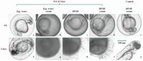
Zebrafish egg bath infection model in various media. Representative embryos were co-incubated with 1 × 10⁶ cells/mL of SC5314 (a-d, f-i) or without C. albicans (control, e, j) in egg water (a, f), egg water/serum (b, g), RPMI medium (c, h), RPMI/serum (d, i) with shaking at 80 rpm and 30°C for 4 h. The embryos were photographed immediately after non-adhered C. albicans cells were removed through washing (a-e) or after an additional 2 days of incubation (f-j). Scale bar = 200 µm. This data are from 3 repeat experiments. Approximately 30 embryos were tested for each treatment. |
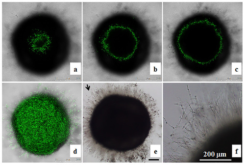
Localization of OG1 Candida albicans cells in zebrafish egg bath infection model. Embryos were co-incubated with 1 × 10⁶ cells/mL of OG1 C. albicans. The representative slices of confocal images (a-c) are shown. The distance between two slices was approximately 55 µm. The whole merged images are presented (d). The phase contrast photos showing C. albicans hyphae were taken by an inverted microscope (e, f). f is the enlargement of the arrow area in e. Scale bars = 200 µm. |
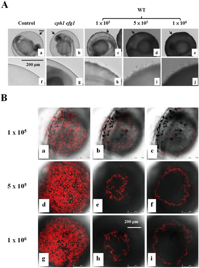
Zebrafish egg bath infection model with different inocula. (A) Embryos were co-incubated in the absence of C. albicans (a, f) or in the presence of 1 × 10⁵ (c, h), 5 × 10⁵ (d-i), or 1 × 10⁶ (e-j) cells/mL of wild-type SC531cells, 1 × 10⁶ (e-j) cells/mL of cph1/cph1 efg1/efg1 mutant cells (b, g) for 4 h. f-j are the enlargement of the arrow areas in a-e. (B) Embryos were co-incubated with 1 × 10⁵ (a-c), 5 × 10⁵ (d-f), or 1 × 10⁶ (g-i) cells/mL of CAF2-dTomato C. albicans. The representative slices (b-c, e-f, h-i) are shown. The distance between two slices was approximately 16 µm. The whole merged images for 1 × 10⁵ (a), 5 × 10⁵ (d) or 1 × 10⁶ (g) cells/mL are presented. Scale bars = 200 µm. |
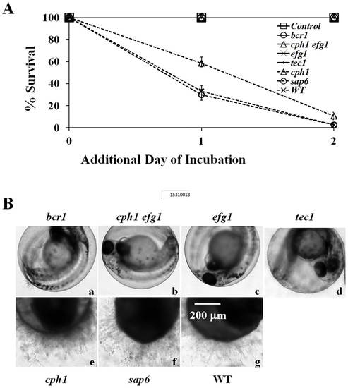
Virulence of C. albicans mutant strains in the infection model. (A) Survival rates of embryos. Embryos alone (Control) or embryos with 5 × 10⁵ cells/mL of bcr1/bcr1, cph1/cph1 efg1/efg1, efg1/efg1, tec1/tec1, cph1/cph1, sap6/sap6, or WT (SC5314) cells in RPMI/serum were incubated at 30°C for 4 h. Survival rates were determined after an additional 1 day and 2 days of incubation. (B) Representative embryos were co-incubated with (a) bcr1/bcr1, (b) cph1/cph1 efg1/efg1, (c) efg1/efg1, (d) tec1/tec1, (e) cph1/cph1, (f) sap6/sap6, or (g) WT (SC5314) cells, and photographed after an additional 2 days of incubation. Scale bar = 200 µm. The data are from 4 repeat experiments. Approximately 70 embryos were tested for each strain. PHENOTYPE:
|