
|
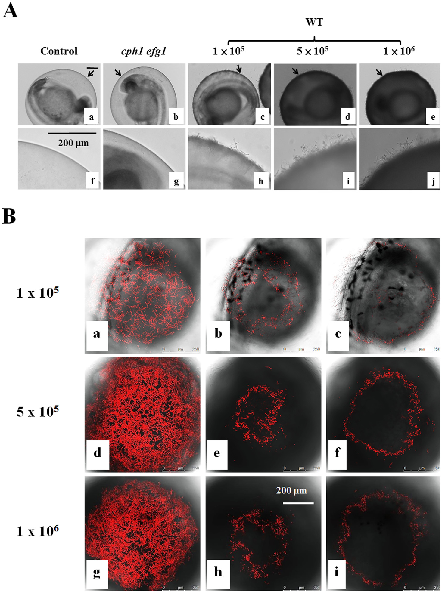
Fig. 3 Zebrafish egg bath infection model with different inocula.
(A) Embryos were co-incubated in the absence of C. albicans (a, f) or in the presence of 1 × 10⁵ (c, h), 5 × 10⁵ (d-i), or 1 × 10⁶ (e-j) cells/mL of wild-type SC531cells, 1 × 10⁶ (e-j) cells/mL of cph1/cph1 efg1/efg1 mutant cells (b, g) for 4 h. f-j are the enlargement of the arrow areas in a-e. (B) Embryos were co-incubated with 1 × 10⁵ (a-c), 5 × 10⁵ (d-f), or 1 × 10⁶ (g-i) cells/mL of CAF2-dTomato C. albicans. The representative slices (b-c, e-f, h-i) are shown. The distance between two slices was approximately 16 µm. The whole merged images for 1 × 10⁵ (a), 5 × 10⁵ (d) or 1 × 10⁶ (g) cells/mL are presented. Scale bars = 200 µm.