
FIGURE
Fig. 2
- ID
- ZDB-FIG-151215-4
- Publication
- Chen et al., 2015 - Zebrafish Egg Infection Model for Studying Candida albicans Adhesion Factors
- Other Figures
- All Figure Page
- Back to All Figure Page
Fig. 2
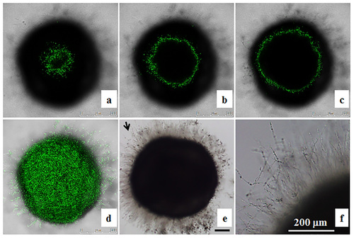
Localization of OG1 Candida albicans cells in zebrafish egg bath infection model. Embryos were co-incubated with 1 × 10⁶ cells/mL of OG1 C. albicans. The representative slices of confocal images (a-c) are shown. The distance between two slices was approximately 55 µm. The whole merged images are presented (d). The phase contrast photos showing C. albicans hyphae were taken by an inverted microscope (e, f). f is the enlargement of the arrow area in e. Scale bars = 200 µm. |
Expression Data
Expression Detail
Antibody Labeling
Phenotype Data
Phenotype Detail
Acknowledgments
This image is the copyrighted work of the attributed author or publisher, and
ZFIN has permission only to display this image to its users.
Additional permissions should be obtained from the applicable author or publisher of the image.
Full text @ PLoS One