
- Title
-
Cellular projections from sensory hair cells form polarity-specific scaffolds during synaptogenesis
- Authors
- Dow, E., Siletti, K., Hudspeth, A.J.
- Source
- Full text @ Genes & Dev.

Hair cells form synapses 2–15 h after mitosis. (A) The fraction of nascent hair cells making contact with afferent terminals (blue) rises briskly following cellular rearrangement. Although the contacts are transitory at the outset, their duration increases progressively until, by 15 h after mitosis, the slope of the line approaches unity, indicating complete stability (red). Time is denoted in hours after mitosis. The gray band marks the period of cellular rearrangement, and the vertical dashed line marks the emergence of projections. N = 30. (B–E) Ribeye-mCherry fusion protein (B,D) and membrane-associated guanylate kinase (MAGUK) (C,E) puncta (arrowheads) appear in late maturity and full maturity hair cells (magenta) but are absent from rearranging cells and early maturity cells (asterisks in B,C). For D and E, P < 0.0001; for D, N = 8, 34, 47, and 176 measurements for the respective hair cell stages; for E, N = 8, 16, 16, and 58 measurements. (F) A time line of hair cell differentiation. The magenta box denotes the extension of projections from nascent hair cells from shortly after their rearrangement until stable synapses have formed (see Figs. 2-4). EXPRESSION / LABELING:
|
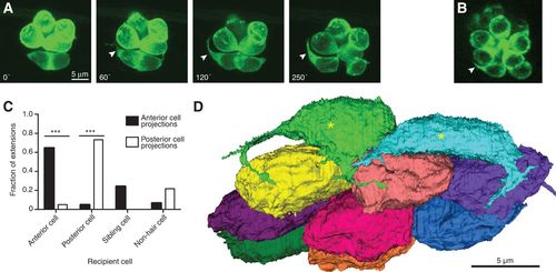
Nascent hair cells extend dynamic basal projections. (A) Time-lapse imaging by fluorescence confocal microscopy of hair cells expressing GFP fused to actin shows the emergence of a basal projection (arrowheads) shortly after the completion of rearrangement. Times are denoted in minutes from the end of rearrangement. (B) A hair cell projection (arrowhead) contains the Map1b-GFP fusion protein (green), indicative of the presence of microtubules. The scale accords with that in A. (C) Projections extend predominantly toward mature hair cells of the same polarity and occasionally toward a sibling cell or a nonhair cell. P < 0.0001 for both group comparisons; N = 9 for anteriorly polarized hair cells, and N = 12 for posteriorly polarized cells. (D) A basal view of an SBEM reconstruction of all of the hair cells in one neuromast shows that early maturity hair cells (asterisks) but not late maturity or full maturity hair cells bear projections. The plane of view is tangential to the larval surface. EXPRESSION / LABELING:
|
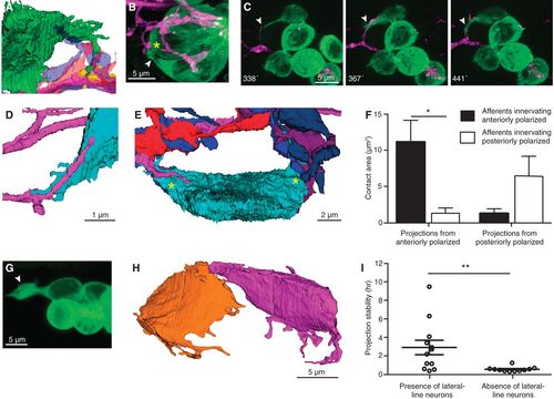
Afferent neurons traverse projections that extend to mature synapses. (A) An SBEM reconstruction shows a hair cell projection (green) extending into an aggregation of variously colored axonal terminals beneath mature ribbon synapses made by another hair cell (yellow). (B) In a frame from a time-lapse sequence, a projection (arrowhead) extends toward a presumptive ribbon synapse (asterisk). (C) A series of time-lapse confocal images shows extension of an afferent terminal along a projection (arrowheads). Times are denoted in minutes after rearrangement. (D) A reconstructed afferent terminal (magenta) traverses a hair cell projection (cyan). (E) An SBEM reconstruction demonstrates that all neurons contact the nascent cell soma on its two projections (asterisks). (F) Projections arising from anteriorly and posteriorly polarized hair cells make the greatest areas of contact with afferent neurons of the same polarity. P < 0.02; N = 4. (G) An unusually large projection (arrowhead) forms in the absence of lateral line neurons. (H) The reconstructed sibling hair cells from a larva whose lateral line ganglion had been ablated exhibit numerous and lengthy projections. (I) In the absence of lateral line neurons, projections show reduced stability. P < 0.01; N = 12 and 11. EXPRESSION / LABELING:
PHENOTYPE:
|
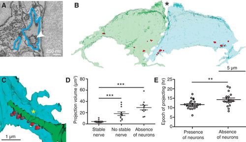
Retraction of projections is associated with stable afferent contact. (A) An SBEM image shows an immature synaptic ribbon (arrowhead) within a hair cell projection (blue outline). (B) In a reconstruction of the sibling hair cells from a recent division, synaptic ribbons (red) cluster at the sites from which projections emerge. N = 10. In this basal view, the partially developed hair bundles are apparent at the cells’ apical ends (asterisk). (C) An afferent terminal of the functionally appropriate subpopulation (green) contacts immature synaptic ribbons (red) on a hair cell projection (blue). A sheet of hair cell membrane (asterisk) folds over the afferent terminal. (D) Enlarged projections are associated with the absence of stable nerve contacts (P < 0.001) or of lateral line neurons (P < 0.0001). N = 10, 12, and 11. (E) The total period during which projections emerge from hair cells increases in the absence of lateral line neurons. P < 0.01; N = 23 and 18. |
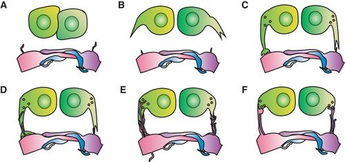
Diagram of a projection-based model of hair cell innervation. (A) During their rearrangement, hair cells make few transient contacts and no stable contacts with afferent terminals. (B) Following the completion of rearrangement, hair cells develop basal projections. (C) The projections extend toward aggregations of afferent neurons at neighboring hair cell synapses. (D) Filopodia from afferent terminals extend along the projections toward the hair cell somata. (E) Afferent filopodia reach the available presynaptic sites at the bases of the projections. (F) Afferent terminals form stable contacts at presynaptic sites, and the hair cell projections retract. |