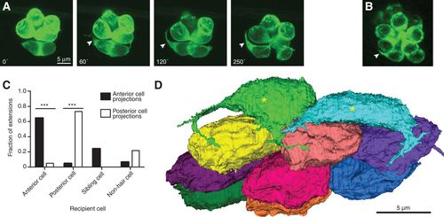
Nascent hair cells extend dynamic basal projections. (A) Time-lapse imaging by fluorescence confocal microscopy of hair cells expressing GFP fused to actin shows the emergence of a basal projection (arrowheads) shortly after the completion of rearrangement. Times are denoted in minutes from the end of rearrangement. (B) A hair cell projection (arrowhead) contains the Map1b-GFP fusion protein (green), indicative of the presence of microtubules. The scale accords with that in A. (C) Projections extend predominantly toward mature hair cells of the same polarity and occasionally toward a sibling cell or a nonhair cell. P < 0.0001 for both group comparisons; N = 9 for anteriorly polarized hair cells, and N = 12 for posteriorly polarized cells. (D) A basal view of an SBEM reconstruction of all of the hair cells in one neuromast shows that early maturity hair cells (asterisks) but not late maturity or full maturity hair cells bear projections. The plane of view is tangential to the larval surface.
|
