
- Title
-
Spindle Assembly Checkpoint Acquisition at the Mid-Blastula Transition
- Authors
- Zhang, M., Kothari, P., Lampson, M.A.
- Source
- Full text @ PLoS One

Pre-MBT embryos lack a functional SAC. Embryos were injected with Alexa 647-Histone H1 and PCNA-GFP proteins, treated with or without nocodazole before the MBT at 2 HPF, then imaged live. Images show cell cycle progression based on Histone H1 and PCNA from an embryo at 2.75 HPF. Insets are displayed with higher contrast settings to show the changing morphology of a single nucleus at different cell cycle stages: I, interphase; M, prometaphase/metaphase; A, anaphase. In the montage, time between metaphase and the next interphase is 8 min for untreated control embryos and 9 min for nocodazole-treated embryos. Graph shows average length of mitosis for each condition (ne14 embryos for each, from three independent experiments). Error bars indicate S.D.; P>0.05; two-tailed Student?s t test; scale bars 20 �m. |
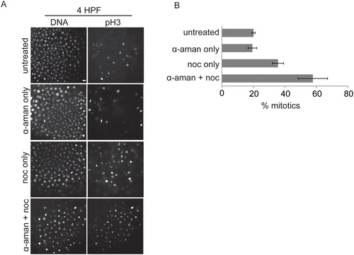
SAC acquisition does not rely on zygotic transcription. 1-cell stage embryos were injected with α-amanitin as indicated, treated with or without nocodazole at 3.25 HPF for 45 min, fixed at 4 HPF, and stained for pH3 and DNA. Representative images are shown (A). The percent of nuclei positive for pH3 was calculated and averaged over multiple embryos (B, ne18 for each condition). Error bars indicate s.e.m, calculated over three independent experiments; scale bar 20 �m. |
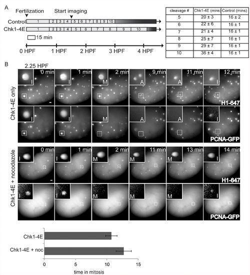
Precocious Chk1 activity and cell cycle elongation are not sufficient for SAC acquisition. (A) Schematic of cell cycle lengths for the first 10 cleavage divisions of embryos injected with Alexa 647-Histone H1, with or without Chk1-4E-GFP mRNA. Cell cycles lengths were measured as time between metaphases, starting from 1.5 HPF, based on Histone H1 morphology. Embryos injected with H1-647 alone (control) had consistent cleavage divisions that each lasted ~16 min as expected [5] while embryos injected with Chk1-4E mRNA and H1-647 had cleavage cycles that progressively lengthened. The measured cell cycle lengths for cleavages 5?10 are indicated by the length of each bar in the schematic and shown in the table (n >4 embryos; error indicates S.D.) (B) Embryos were injected with Alexa 647-Histone H1 and PCNA-GFP proteins and Chk1-4E mRNA, treated with our without nocodazole at 2.25 HPF, then imaged live until 3 HPF. Images show cell cycle progression based on Histone H1 and PCNA. Insets are displayed with higher contrast settings to show nuclear morphology as in Fig. 1. In the montage, time between metaphase and the next interphase is 10 min without nocodazole and 12 min for nocodazole-treated embryos. There was no measurable progressive lengthening of mitoses during the cleavage divisions, regardless of drug treatment. Graph shows average length of mitosis for each condition (ne12 for each condition, from three independent experiments). Error bars indicate S.D.; P>0.05; two-tailed Student?s t test; scale bars 20 �m. |
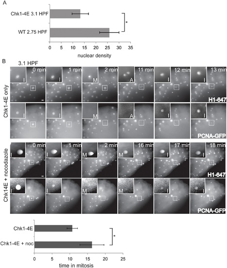
SAC acquisition is independent of the N:C ratio. (A) Nuclear density was measured for embryos injected with Alexa 647-Histone H1 and PCNA-GFP proteins and Chk1-4E mRNA at 3.1 HPF, or embryos injected only with H1-647 and PCNA-GFP proteins at 2.75 HPF. Error bars are S.D.; ne11; Pd0.001. (B) Embryos were injected with Alexa 647-Histone H1 and PCNA-GFP proteins and Chk1-4E mRNA, treated with or without nocodazole at 3.1 HPF, then imaged live. Images show cell cycle progression based on Histone H1 and PCNA-GFP. Insets are displayed with higher contrast settings to show nuclear morphology as in Fig. 1. In the montage, time between metaphase and the next interphase is 10 min without nocodazole and 16 min for nocodazole-treated embryos. Graph shows average length of mitosis for each condition (ne13 from four independent experiments). Error bars indicate S.D.; * Pd 0.001; two-tailed Student?s t test; scale bars 20 �m. |