
- Title
-
Coral-Derived Compound WA-25 Inhibits Angiogenesis by Attenuating the VEGF/VEGFR2 Signaling Pathway
- Authors
- Lin, S.W., Huang, S.C., Kuo, H.M., Chen, C.H., Ma, Y.L., Chu, T.H., Bee, Y.S., Wang, E.M., Wu, C.Y., Sung, P.J., Wen, Z.H., Wu, D.C., Sheu, J.H., Tai, M.H.
- Source
- Full text @ Mar. Drugs
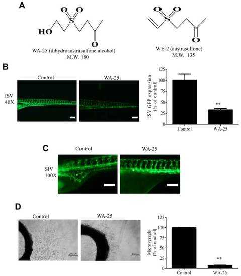
Angiogenesis inhibition by WA-25 in vivo and ex vivo (A) Chemical structures of WA-25 (dihydroaustrasulfone alcohol) and WE-2 (austrasulfone); (B) Effect of WA-25 on intersegmental vessels (ISVs) development in transgenic Tg(fli-1:EGFP)y1 zebrafish embryos. (Left panel) Representative photographs of ISV in control and WA-25 (50 µM)-treated zebrafish at 48 h poat-fertilization (hpf). Magnification 40×; scale bar, 50 µm. (Right panel) Quantification analysis of enhanced green fluorescent protein (EGFP) intensities in the ISVs in WA-25 (50 µM)-treated embryos. Data are represented as mean ± SEM (n = 12); (C) Effect of WA-25 (50 µM) on SIV development in transgenic Tg(fli-1:EGFP)y1 zebrafish embryos. Photographs of control and WA-25-treated zebrafish were taken at 72 hpf. Magnification, 100×; scale bar, 100 µm. Asterisks indicate arcades in the vesicle-like structure (D) Effect of WA-25 on microvessel sprouting in aortic rings. Rat aortic rings were placed in Matrigel and treated with WA-25 (20 µM) for 7 days. Scale bar, 2 mm. Data are represented as mean ± SEM (n = 12). ** p < 0.01. |
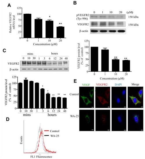
Effect of WA-25 on VEGFR2 expression in HUVECs. HUVECs were treated with WA-25 (1–20 µM) for 48 h and separately subjected to VEGFR2 mRNA and protein expression assay. (A) VEGFR2 mRNA level was determined using quantitative qRT-PCR analysis. (B) Dose-dependent (48 h) and (C) time-dependent effect of WA-25 on VEGFR2 protein expression was measured using Western blot analysis (D) Flow cytometric analysis of surface VEGFR2 expression after WA-25 (20 µM) treatment for 48 h (E) VEGF and VEGFR2 protein expressions were analyzed using immunofluorescence. After WA-25 (20 µM) treatment for 48 h, the cell surface VEGFR2 expression in endothelial cells was analyzed using FACScan. Data are represented as mean ± SD in triplicates. * p < 0.05; ** p < 0.01, versus control groups. |
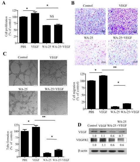
Effects of exogenous VEGF on WA-25-induced neovascularization blockade in vitro. After treatment with WA-25 (20 µM) with or without VEGF (10 ng/mL), the effects of WA-25 on VEGF-induced proliferation (A); migration (B); and tube formation (C) were determined in HUVECs; (D) Immunoblot analysis of VEGF and VEGFR2 expression in HUVECs after treatment with WA-25 (20 µM) with or without VEGF (10 ng/mL) for 24 h. Data are represented as mean ± SD of quadruplicate experiments. Asterisks indicate statistical significance versus control (* p < 0.05 and ** p < 0.01). NS, not significant. |

Effect of VEGF-A supply on WA-25-induced neovascularization blockade in vivo (A) Effect of exogenous VEGF-A on WA-25-induced angiogenesis blockade on the microvessel sprouting in aorta rings. Rat aortic rings were placed in Matrigel and treated with VEGF-A or WA-25. The effect of WA-25 on the formation of vessel sprouts from various aortic samples was observed on Day 7; (B) Effect of exogenous VEGF-A on WA-25-induced angiogenesis in Tg(kdrl:mCherryci5-fli1a:negfp)y7 zebrafish embryos. Embryos were treated with WA-25 (50 µM) at 6 hpf with or without VEGF (10 ng/mL) and then imaged at 24 hpf. The number of endothelial cells on the ISV was determined by counting the green nuclei over the red blood vessel. Data are represented as mean ± SD (n = 12). Asterisks indicate statistical significance versus control (* p < 0.05 and ** p < 0.01). |