
- Title
-
Mutations in KATNB1 Cause Complex Cerebral Malformations by Disrupting Asymmetrically Dividing Neural Progenitors
- Authors
- Mishra-Gorur, K., Çağlayan, A.O., Schaffer, A.E., Chabu, C., Henegariu, O., Vonhoff, F., Akgümüş, G.T., Nishimura, S., Han, W., Tu, S., Baran, B., Gümüş, H., Dilber, C., Zaki, M.S., Hossni, H.A., Rivière, J., Kayserili, H., Spencer, E.G., Rosti, R.Ö., Schroth, J., Per, H., Çağlar, C., Çağlar, Ç., Dölen, D., Baranoski, J.F., Kumandaş, S., Minja, F.J., Erson-Omay, E.Z., Mane, S.M., Lifton, R.P., Xu, T., Keshishian, H., Dobyns, W.B., Chi, N.C., Šestan, N., Louvi, A., Bilgüvar, K., Yasuno, K., Gleeson, J.G., Günel, M.
- Source
- Full text @ Neuron
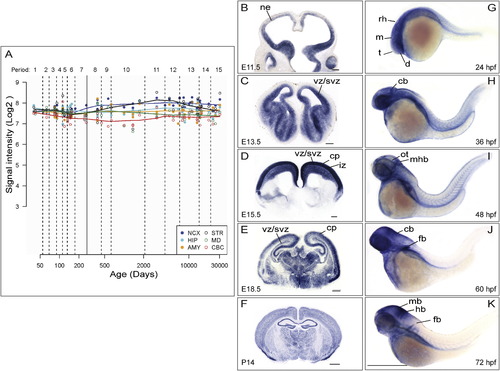
KATNB1 Is Highly Expressed in the Developing Brain (A) KATNB1 is expressed across all regions and developmental periods in the human brain. KATNB1 exon array signal intensity. NCX, neocortex; STR, striatum; HIP, hippocampus; MD, mediodorsal nucleus of the thalamus; AMY, amygdala; CBC, cerebellar cortex. In developing mouse brain, Katnb1 is expressed in neural progenitors until midneurogenesis (E11.5, E13.5) (B and C), and then is also expressed in postmitotic neurons in the cortical plate (E15.5, E18.5) (D and E), and widespread expression was observed in postnatal brain (P14) (F). ne, neuropithelium; vz, ventricular zone; svz, subventricular zone; iz, intermediate zone; cp, cortical plate. Scale bars (G–I), 200 µm, (J and K), 500 µm. Similarly, katnb1 is expressed in the brain in the developing zebrafish embryo (G–K). Lateral views of whole-mount in situ hybridization of the brain and torso of zebrafish embryos reveal the expression pattern of katnb1 at 24 hr postfertilization (hpf) (G), 36 hpf (H), 48 hpf (I), 60 hpf (J), and 72 hpf (K). During early developmental stages (G and H), katnb1 mRNA expression is ubiquitous throughout the embryo, including the cephalic region. As the embryos develop further (I–K), katnb1 mRNA expression becomes restricted to neural tissue. Black lines point to various anatomical structures. d, diencephalon; t, telencephalon; m, mesencephalon; rh, rhombomeres; cb, cerebellum; ot, optic tectum; mhb, midbrain hindbrain boundary; mb, midbrain; hb, hindbrain; fb, fin bud. Scale bar, 500 µm.EXPRESSION / LABELING:
|
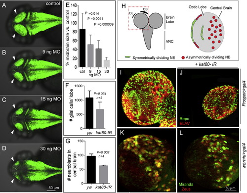
Knockdown of KATNB1 Orthologs in Zebrafish and Drosophila Results in Small Brain Phenotype katnb1 morpholino reduces zebrafish midbrain size. Confocal microscopy shows that the katnb1 morphants at (B) 9 nanograms (ng), (C) 15 ng, and (D) 30 ng have smaller midbrains (arrows) as compared with (A) control at 2 days postfertilization (dpf). The reduction in brain size is statistically significant (E). Zebrafish brain is labeled with green fluorescence by Tg (HuC:Kaede). (H) Left panel: schematic of the Drosophila brain; box indicates brain lobe imaged. Right panel: schematic of a single brain lobe marks the location of symmetrically dividing neuroepithelium (NE; green) and asymmetrically dividing neuroblasts (NBs; red). (I and J) Expression of kat80-IR with prospero-Gal4 results in a dramatically reduced brain size in third instar larvae. There is an overall reduction in the number of neurons and glia generated as seen by ELAV (red) and Repo (green) staining, respectively. (K and L) kat80-IR expressed under worniu-Gal4, UAS-mir::GFP, UAS-zeus::mCherry results in a significant reduction in NB number in central brain. Images in (K) and (L) are 3D projections of identical Z-sections. (F) Quantification of glial cell counts seen in (I) and (J). There is significantly reduced number of glial cells in kat80-IR larvae (error bars indicate SD; yw, 1,080 ± 110; kat80-IR, 673 ± 116; two-tailed t test, p = 0.034). (G) GFP- and RFP-positive cells were quantified using 3D projections of identical Z-stacks from worniu>gal4 and worniu>kat80-IR brains, which reveal a significant reduction in central brain NBs per brain lobe (yw, 96.5 ± 7.9; kat80-IR, 62.5 ± 2.3; p = 0.002) (see also Figure S3). PHENOTYPE:
|