
IMAGE
Fig. 4
- ID
- ZDB-IMAGE-150320-2
- Genes
- Publication
- Mishra-Gorur et al., 2014 - Mutations in KATNB1 Cause Complex Cerebral Malformations by Disrupting Asymmetrically Dividing Neural Progenitors
- All Figures
- Figures for Mishra-Gorur et al., 2014
Image
|
Figure Caption
Fig. 4
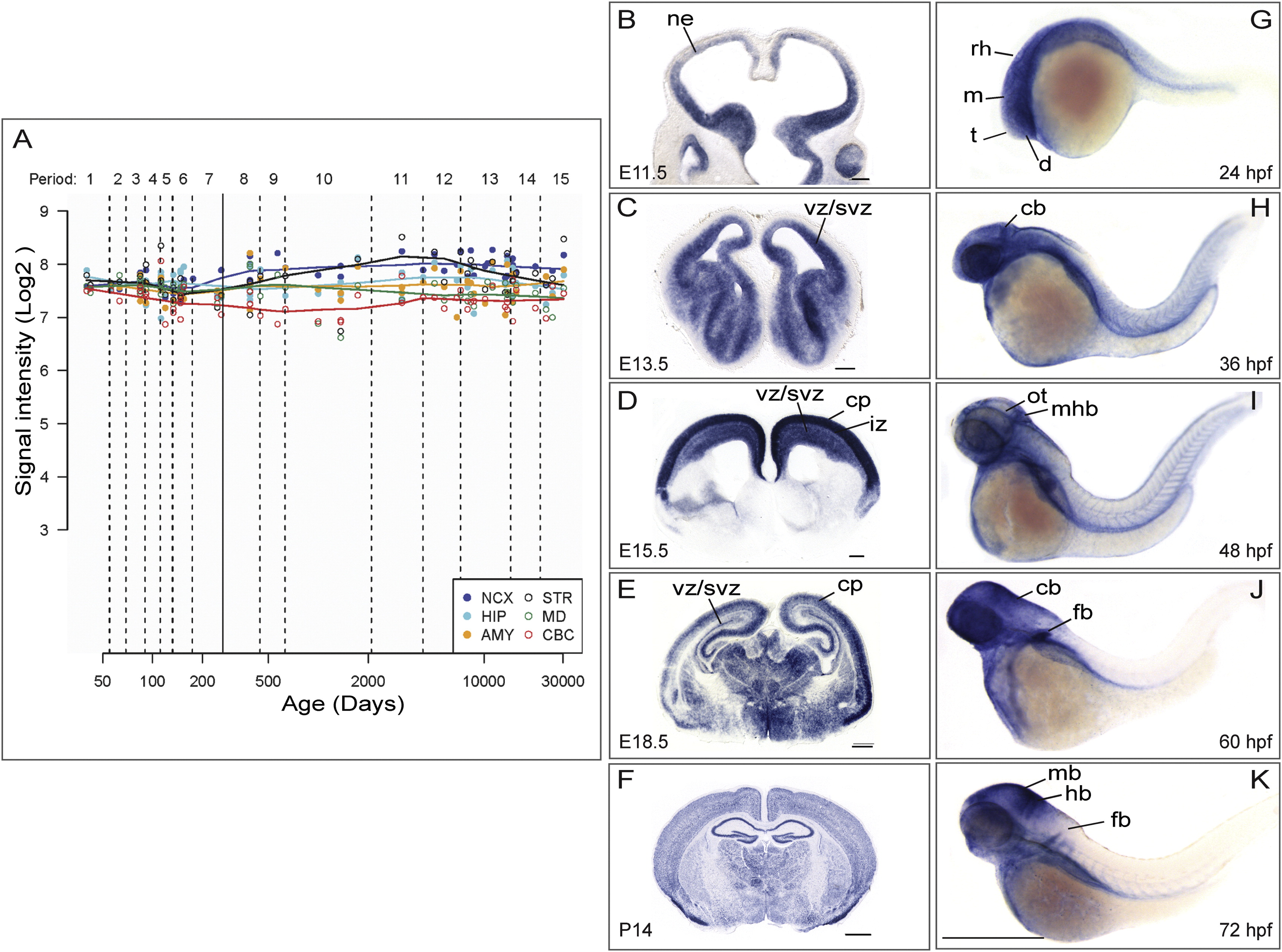
KATNB1 Is Highly Expressed in the Developing Brain
(A) KATNB1 is expressed across all regions and developmental periods in the human brain. KATNB1 exon array signal intensity. NCX, neocortex; STR, striatum; HIP, hippocampus; MD, mediodorsal nucleus of the thalamus; AMY, amygdala; CBC, cerebellar cortex. In developing mouse brain, Katnb1 is expressed in neural progenitors until midneurogenesis (E11.5, E13.5) (B and C), and then is also expressed in postmitotic neurons in the cortical plate (E15.5, E18.5) (D and E), and widespread expression was observed in postnatal brain (P14) (F). ne, neuropithelium; vz, ventricular zone; svz, subventricular zone; iz, intermediate zone; cp, cortical plate. Scale bars (G–I), 200 µm, (J and K), 500 µm. Similarly, katnb1 is expressed in the brain in the developing zebrafish embryo (G–K). Lateral views of whole-mount in situ hybridization of the brain and torso of zebrafish embryos reveal the expression pattern of katnb1 at 24 hr postfertilization (hpf) (G), 36 hpf (H), 48 hpf (I), 60 hpf (J), and 72 hpf (K). During early developmental stages (G and H), katnb1 mRNA expression is ubiquitous throughout the embryo, including the cephalic region. As the embryos develop further (I–K), katnb1 mRNA expression becomes restricted to neural tissue. Black lines point to various anatomical structures. d, diencephalon; t, telencephalon; m, mesencephalon; rh, rhombomeres; cb, cerebellum; ot, optic tectum; mhb, midbrain hindbrain boundary; mb, midbrain; hb, hindbrain; fb, fin bud. Scale bar, 500 µm.
Figure Data
Acknowledgments
This image is the copyrighted work of the attributed author or publisher, and
ZFIN has permission only to display this image to its users.
Additional permissions should be obtained from the applicable author or publisher of the image.
Full text @ Neuron