- Title
-
Role of mef2ca in developmental buffering of the zebrafish larval hyoid dermal skeleton
- Authors
- Delaurier, A., Huycke, T.R., Nichols, J.T., Swartz, M.E., Larsen, A., Walker, C., Dowd, J., Pan, L., Moens, C.B., and Kimmel, C.B.
- Source
- Full text @ Dev. Biol.
|
The shapes of the opercle (Op) – branchiostegal ray 3 (BR) bones are outstandingly diverse in mef2ca mutants. Confocal projections (dorsal up, anterior to the left), bones vitally stained with Alizarin Red and live-imaged at 6 dpf. (A and B) wild type (WT), with (B) evidently the more advanced (h: hyomandibula, i: interopercle rudiment, and 2: a second BR). (C–J) mef2ca mutants. The expanded mutant bone in (C) appears to a mirror-image duplicated Op (Op2), and a strut (s) of bone may, or may not, include a BR rudiment. Examples of mutants in (F–J) include the BR bridged to the OP (b, panel J). About 2/3 of the mutants in the single pair family used for this and Fig. 2 include either a strut or bridge as part of the ectopic bone formation. Scale bar 50 μm. PHENOTYPE:
|
|
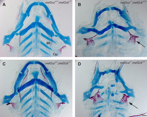
mef2cb may not function in OpBR development, but functions partially redundantly with mef2ca in cartilage development. Ventral views of dissected flat mounts at 6 dpf, stained with Alcian Blue and Alizarin Red. (A and C) The skeletons (bone, red; cartilage, blue) appear phenotypically normal. (B and C) The OpBR bones show the expansion phenotype (arrows), the cartilages are disrupted, substantially more so in (D) than (B). Scale bar 50 μm. PHENOTYPE:
|
|
Loss of mef2ca results in the ectopic differentiation of cells with Op joint identity. Confocal projections of larvae expressing trps1:EGFP at 6 dpf, live imaged after counterstaining vitally with Alizarin Red. (A) Wild-type cells of the Op-hyosymplectic articulation, but not BR-ceratohyal cartilage articulation, express high levels of trps1:EGFP (arrow). (B) Mutant cells of the expanded (transformed) BR-ceratohyal articulation now show high levels of trps1:EGFP (arrow). (C) A region of ectopic bone cells not involving the BR, express high levels of trps1:EGFP (arrow). A stunted bony spur at the position of the BR does not show high expression (asterisk). (D) A mutant showing less ectopic bone expansion matches the wild type (A) in trps1:EGFP expression. Scale bar 50 μm. |
|
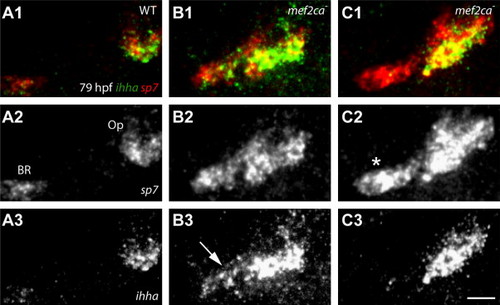
sp7-expressng osteoblasts of the Op and ectopic Op2 specifically express ihha during phase 2 of Op development. Confocal projections of larvae labeled for ihha and sp7, RNA in situ hybridization. (A) ihha is expressed at high levels in the wild-type Op, but only minimally in the BR. (B) Expanded ihha mef2ca mutant expression is present at the location of the BR (B3, arrow). (C) Op ihha expression is expanded (C3, arrow), but not detected at the location of the BR (C2, asterisk). Scale bar 50 μm. EXPRESSION / LABELING:
|
|
Variation in the expression of dlx5:EGFP in preskeletal condensations (3 dpf, upper panels) does not predict later variation in the larval mutant OpBR skeletal elements (re-imaged at 6 dpf, lower panels) of mef2ca mutants. Live preparations, bone vitally counterstained with Alizarin Red. At the early stage the wild type shows two fairly well separated expression domains (A1, six animals imaged at 3 dpf, and then again at 6 dpf). These domains are merged together in mef2ca mutants that later show different OpBR expansion phenotypes (B1–D1, eight mutants imaged). Scale bar 50 μm. EXPRESSION / LABELING:
PHENOTYPE:
|
|
Differences in position and timing of ectopic osteoblast appearance and outgrowth explains skeletal phenotypic variation. (A1–C4) Confocal projections from time lapse recordings starting at 55 hpf. sp7:EGFP labeling. (A5–C5) The same larvae at 6 dpf, bone matrix counterstained with Alizarin Red. (A) In the wild type a small collective of sp7:EGFP-expressing osteoblasts emerge (A1), and then osteoblasts are newly recruited at the posterior-ventral apex (A2, arrow) to expand the bone linearly (A2–A4). (B) A mef2ca mutant in which ectopic osteoblasts are added to the anterior side of the Op axis (B2). The outgrowth expands with new osteoblast addition in both the orthogonal (B3, arrow) and parallel pattern (B4, arrow). At 6 dpf the Op appears duplicated; a BR is absent (B5). (C) A mef2ca mutant in which two groups of ectopic osteoblasts arise (C2 and C4, arrows) and then merge. At 6 dpf the Op has a short spur, and appears duplicated. Scale bar 25 μm (A1–C4) and 50 μm (A5–C5). |
Reprinted from Developmental Biology, 385(2), Delaurier, A., Huycke, T.R., Nichols, J.T., Swartz, M.E., Larsen, A., Walker, C., Dowd, J., Pan, L., Moens, C.B., and Kimmel, C.B., Role of mef2ca in developmental buffering of the zebrafish larval hyoid dermal skeleton, 189-99, Copyright (2014) with permission from Elsevier. Full text @ Dev. Biol.


