
- Title
-
Zebrafish guanylate cyclase type 3 signaling in cone photoreceptors
- Authors
- Fries, R., Scholten, A., Säftel, W., and Koch, K.W.
- Source
- Full text @ PLoS One
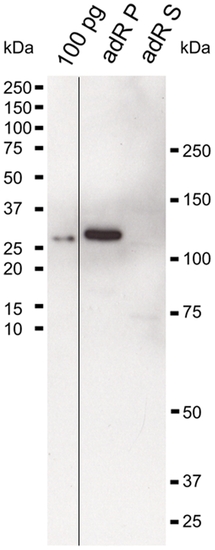
Specificity of anti-zGC3 antibody. Hundred pg of the antigen, the pellet adRP) and soluble fraction (adRS) of 1/10 of an adult zebrafish retina each were probed with the purified polyclonal anti-zGC3 antibody (1:1000); secondary antibody was a goat anti rabbit coupled to peroxidase (1:5000). EXPRESSION / LABELING:
|
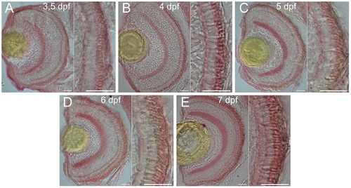
Temporal expression profile of zGC3 during larval development. Cryosections of larval eyes were probed with the anti-zGC3 antibody (1:400) at 3.5 – 7 dpf (A–E) and stained with Fast Red. Left part of each panel is a section through the whole eye, right part shows the photoreceptor layer. Scale bar = 20 μm. EXPRESSION / LABELING:
|
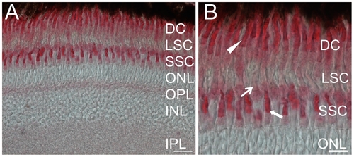
Immunostaining of zGC3 in cryosections of adult zebrafish retina. Sectionas were probed with the anti-zGC3 antibody (1:400). The secondary antibody was used at 1:200 dilution. (A) Section of a whole retina, cell layers are indicated as double cones (DC), long single cones (LSC), short single cones (SSC), outer nuclear layer (ONL), outer plexiform layer (OPL), inner nuclear layer (INL) and inner plexiform layer (IPL). (B) Magnified part of (A). DC: closed arrowhead; LSC: thin arrow; SSC: bold arrow. Scale bar = 20 μm in (A) and 10 μM in (B). EXPRESSION / LABELING:
|
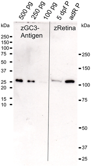
Quantitative estimation of zGC3 in adult zebrafish retina by western blotting. Different amounts of zGC3 antigen (100 pg, 250 pg and 500 pg) were used as calibration standards and compared with the pellet fraction of 5 dpf old larva eyes (10 eyes were used for homogenization) and of 1/10 of an adult retina (adRP). |
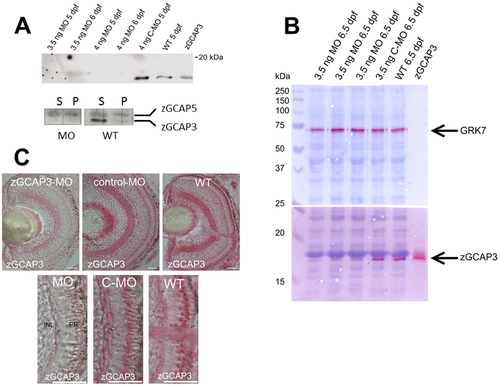
Morpholino knockdown of zGCAP3 in zebrafish larvae. (A) upper panel: Immunoblotting of homogenized eyes obtained from larvae at 5 and 6 dpf that had been injected with the indicated amount of MOs. Recombinant nonmyristoylated zGCAP3 (1 ng) was loaded on the very right lane. Purified polyclonal anti-zGCAP3 was used at 1:250, second antibody was a peroxidase-coupled goat anti-rabbit IgG at a dilution of 1:5000. Lower panel: Immunoblotting as above of MO treated and WT larvae (5 dpf, 10 eyes each). Supernatant (S) and pellet (P) fraction of larval eye homogenates were probed by polyclonal anti-zGCAP3 and anti-zGCAP5 antibodies (dilution of 1:200 and 1:250, respectively). Second antibody was a peroxidase-coupled goat anti-rabbit IgG at a dilution of 1:2500. (B) Analysis of larval eyes at 6.5 dpf by immunoblotting. The same larvae (three lanes that were identically labeled “MO 6.5 dpf”) had been investigated in the optomotor response measurements at 6 dpf. Proteins were first fixed in the polyacrylamide gel according to ref. [34] before transfer to the blot membrane. Reactive antibody binding was visualized by using Fast Red. The anti-GRK7 antibody was used at a dilution of 1:1000. Use of other antibodies as in (A). (C) Immunohistochemistry of larval eyes at 5 dpf as indicated. Cryosection of 10 µm were labeled with the purified polyclonal anti-zGCAP3 antibody at a dilution of 1:2000, secondary antibody was a goat anti-rabbit conjugated to alkaline phosphatase, dilution at 1:200. Scale bars: 20 µm. The lower part of the figure is a magnified part of the upper figures showing the photoreceptor cell layer. |
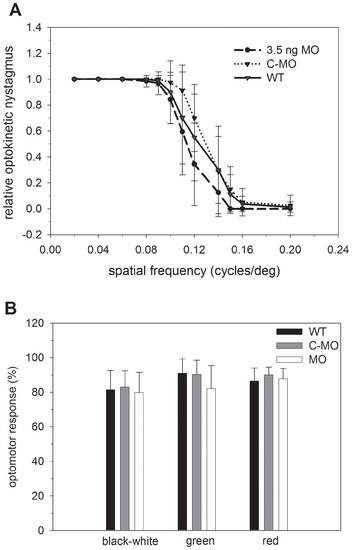
Visual behavioral assays. (A) Optokinetic response measurements of WT, MO and control MO larvae: 8-10 larvae at 5 dpf were investigated by presenting stimuli of a pattern of black and white stripes as described in the Methods. Results of black and green, black and red, and black and blue striped pattern are shown in supplementary Fig. S1. The relative optokinetic nystagmus is shown as a function of the spatial frequency (mean±s.d.). (B) Optomotor response evaluation of larvae as indicated at 6 dpf. For the black and white stimulus the mean±s.d. of 52 measurements is shown (MO versus WT: t = 0.98, Pe0.05; MO versus control MO: t = 2.0, P = 0.05; control MO versus WT: t = 1.05, P≥0.05), for the coloured stimuli the mean±s.d. of 12 data sets each is shown (black/green MO versus WT: t = 1.94, P e 0.05; MO versus control MO: t = 1.8, P≥0.05; control MO versus WT: t = 0.19, P≥0.05). Black / red: MO versus WT: t = 0.44, P≥0.05; MO versus control MO: t = 1.05, P≥0.05; control MO versus WT: t = 1.4, P≥0.05). PHENOTYPE:
|