
Fig. 6
- ID
- ZDB-FIG-131003-5
- Publication
- Fries et al., 2013 - Zebrafish guanylate cyclase type 3 signaling in cone photoreceptors
- Other Figures
- All Figure Page
- Back to All Figure Page
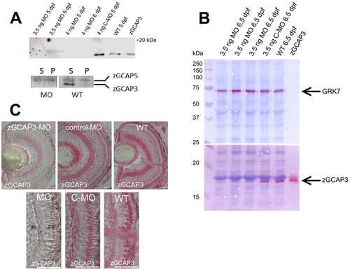
Morpholino knockdown of zGCAP3 in zebrafish larvae. (A) upper panel: Immunoblotting of homogenized eyes obtained from larvae at 5 and 6 dpf that had been injected with the indicated amount of MOs. Recombinant nonmyristoylated zGCAP3 (1 ng) was loaded on the very right lane. Purified polyclonal anti-zGCAP3 was used at 1:250, second antibody was a peroxidase-coupled goat anti-rabbit IgG at a dilution of 1:5000. Lower panel: Immunoblotting as above of MO treated and WT larvae (5 dpf, 10 eyes each). Supernatant (S) and pellet (P) fraction of larval eye homogenates were probed by polyclonal anti-zGCAP3 and anti-zGCAP5 antibodies (dilution of 1:200 and 1:250, respectively). Second antibody was a peroxidase-coupled goat anti-rabbit IgG at a dilution of 1:2500. (B) Analysis of larval eyes at 6.5 dpf by immunoblotting. The same larvae (three lanes that were identically labeled “MO 6.5 dpf”) had been investigated in the optomotor response measurements at 6 dpf. Proteins were first fixed in the polyacrylamide gel according to ref. [34] before transfer to the blot membrane. Reactive antibody binding was visualized by using Fast Red. The anti-GRK7 antibody was used at a dilution of 1:1000. Use of other antibodies as in (A). (C) Immunohistochemistry of larval eyes at 5 dpf as indicated. Cryosection of 10 µm were labeled with the purified polyclonal anti-zGCAP3 antibody at a dilution of 1:2000, secondary antibody was a goat anti-rabbit conjugated to alkaline phosphatase, dilution at 1:200. Scale bars: 20 µm. The lower part of the figure is a magnified part of the upper figures showing the photoreceptor cell layer. |
| Genes: | |
|---|---|
| Antibodies: | |
| Fish: | |
| Knockdown Reagent: | |
| Anatomical Term: | |
| Stage Range: | Day 5 to Day 6 |