
- Title
-
HuB (elavl2) mRNA Is Restricted to the Germ Cells by Post-Transcriptional Mechanisms including Stabilisation of the Message by DAZL
- Authors
- Wiszniak, S.E., Dredge, B.K., and Jensen, K.B.
- Source
- Full text @ PLoS One
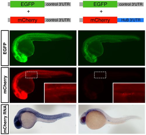
Post-transcriptional regulation of the HuB 32UTR restricts HuB expression to the germ cells. Synthetic reporter RNAs, indicated schematically at top of figure, were transcribed in vitro and 200 pg of both the EGFP and mCherry reporter RNAs were microinjected into 1-cell stage embryos, which were then imaged at approximately 24 hpf. All EGFP images were taken at 2000 ms exposure time, and all mCherry images were taken at 150 ms exposure time. Top panels: EGFP protein expression; Middle panels: mCherry protein expression, insets show a higher magnification image of the germ cells (the dashed box indicates the position of the higher magnification image); Bottom panel: whole-mount in situ hybridisation using an antisense probe directed against the mCherry coding sequence. |
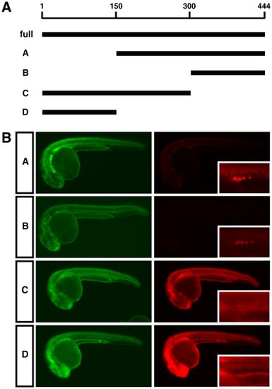
The last 144 nt of the HuB 32UTR are necessary and sufficient for post-transcriptional regulation of HuB mRNA. (A) Schematic representation of the four HuB 32UTR deletion reporter RNAs constructed, termed A, B, C and D. The 32UTR regions present in each of the reporters are indicated by black bars, and are shown relative to the full-length 32UTR of 444 nucleotides at the top. (B) Expression of EGFP and mCherry protein in embryos injected with the indicated deletion mutant reporters. 200 pg of each reporter was injected. All EGFP images were taken at 2000 ms, and all mCherry images were taken at 150 ms. Higher magnification images of mCherry expression in the germ cells are shown as insets. |
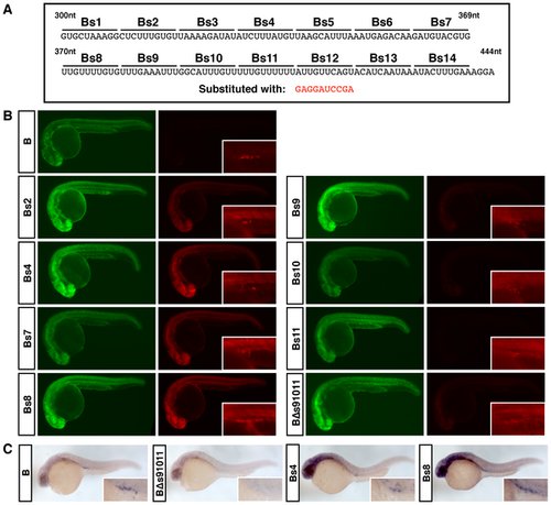
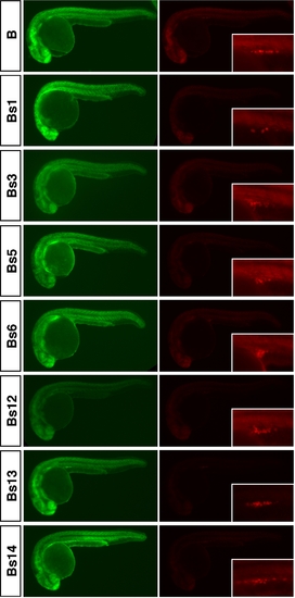
Cis-regulatory elements control differential HuB mRNA stability in the soma and germ cells. (A) 32UTR sequence of the B deletion reporter. Base substitutions were made in 10 nucleotide blocks to create the B substitution mutants Bs1–Bs14. Each 10 nucleotide block was substituted with the sequence GAGGATCCGA. (B) Expression of EGFP and mCherry protein in embryos injected with the indicated Bs substitution mutant reporters. 200 pg of each reporter was injected. All EGFP images were taken at 2000 ms, and all mCherry images were taken at 150 ms. Higher magnification images of mCherry expression in the germ cells are shown as insets. (C) Whole-mount in situ hybridisation using an antisense probe directed against the mCherry coding sequence. Higher magnification images of germ cells are shown as insets. |
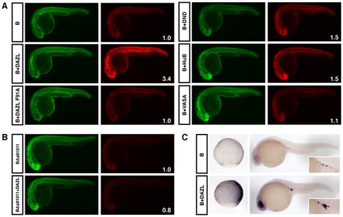
DAZL protein is able to stabilise the HuB mRNA. (A) Expression of EGFP and mCherry protein in embryos injected with 150 pg each of EGFP and mCherry-B reporter RNAs, either alone (labelled B), or with ubiquitous overexpression of HA-tagged DAZL, DND, HuB (400 amol RNA) or VASA (200 amol RNA) as labelled. All EGFP images were taken at 2000 ms and all mCherry images were taken at 500 ms. The fold change in the somatic expression of mCherry from the B reporter-protein combination compared to the B reporter alone is indicated as an inset number in white. (B) mCherry-BΔs91011 reporter RNA +/- overexpression of DAZL. (C) Whole-mount in situ hybridisation using an antisense probe directed against the mCherry coding sequence. Embryos at 9 hpf are shown in the left panels (sagittal view with dorsal side to the right), and embryos at 24 hpf are shown in the right panels with germ cells shown as insets. |
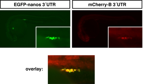
The B 32UTR reporter is expressed in germ cells. Synthetic reporter RNAs were transcribed in vitro and both the EGFP-nanos and mCherry-B reporter RNAs were microinjected into 1-cell stage embryos, which were then imaged at approximately 24 hpf. Top panels: EGFP and mCherry protein expression; insets show a higher magnification image of the germ cells; Bottom panel: image overlays of both EGFP and mCherry protein expression showing coincident expression; image intensity of the mCherry expression was increased to allow for visualisation of protein co-expression in yellow. |
Bs substitution reporters that show no significant differences relative to the parental B reporter. Expression of EGFP and mCherry protein in embryos injected with the indicated Bs substitution mutant reporters. 200 pg of each reporter was injected. All EGFP images were taken at 2000 ms, and all mCherry images were taken at 150 ms. Higher magnification images of mCherry expression in the germ cells are shown as insets. |