- Title
-
Cse1l Is a Negative Regulator of CFTR-Dependent Fluid Secretion
- Authors
- Bagnat, M., Navis, A., Herbstreith, S., Brand-Arzamendi, K., Curado, S., Gabriel, S., Mostov, K., Huisken, J., and Stainier, D.Y.
- Source
- Full text @ Curr. Biol.
|
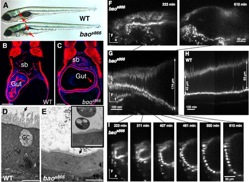
baos866 Mutants Undergo a Dramatic and Rapid Expansion of the Gut Lumen between 96 and 120 Hours Postfertilization (A) Bright-field image of 120 hr postfertilization (hpf) wild-type (WT) and baos866 mutant larvae. Arrows point to the edges of the gut lumen. (B and C) Confocal images of cross-sections of 120 hpf WT (B) and baos866 mutant (C) larvae. The arrow in (C) points to a delaminating cell. Red is F-actin and blue is DAPI; sb indicates swim bladder. Scale bars represent 50 μm. (D and E) Dramatic shortening of microvilli (arrows) in baos866 mutant enterocytes observed by transmission electron microscopy at 120 hpf. Inset in (E) shows remnants of apoptotic cells found in the lumen. GC indicates goblet cell. Scale bars represent 10 μm. (F–I) Rapid expansion of the gut lumen in baos866 mutants expressing histone 2A:GFP. (F) Still images (lateral views) from a baos866 mutant selective plane illumination microscopy (SPIM) recording between 96 and 120 hpf showing first and last frames. (G and H) Kymographs from baos866 mutant (G) and WT (H) gut. Lumen expansion in the mutant occurred in ~200 min; no cell division was observed. (I) Still images (lateral views) from the baos866 mutant SPIM recording corresponding to the kymograph shown in (G). |
|
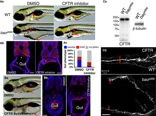
Lumen Expansion in baos866 Mutants Results from Increased CFTR-Dependent Fluid Secretion (A) Inhibition of CFTR blocks lumen expansion in baos866 mutants. (Aa) Bright-field images of WT and baos866 mutants incubated with DMSO (0.1%) or the CFTR inhibitor T08 (5 μM) from 72 to 120 hpf. Arrows point to the edges of the gut lumen. (Ab) Confocal images of cross-sections of 144 hpf control and T08-treated baos866 mutants. Arrowheads point to delaminating cells. Red is F-actin and blue is DAPI. Scale bars represent 50 μm. (Ac) Quantification of the gut phenotype in control and T08-treated baos866 mutants. Larvae were placed in three phenotypic categories (no phenotype, mild phenotype, or severe phenotype) and then genotyped. (B) Activation of CFTR in WT phenocopies the gut lumen expansion defect of baos866 mutants. (Ba) Bright-field image of 144 hpf WT larva treated with DMSO (0.15%) or CFTR-Act9 (15 μM). Arrows point to the edges of the gut lumen. (Bb) Confocal images of cross-sections of 144 hpf control and CFTR-Act9-treated WT larvae. Red is F-actin and blue is DAPI. Scale bars represent 20 μm. (C) Cftr expression and localization is not affected in baos866 mutants compared to WT. (Ca) Immunoblot of 120 hpf WT and baos866 mutants probed against Cftr and β-tubulin. (Cb) Confocal images of whole mounts of 120 hpf WT (top) and baos866 mutant (bottom) stained for Cftr. Arrows point to the apical surface of the gut. Anterior is to the right. Scale bars represent 50 μm. |
|
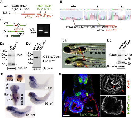
Isolation of the bao Gene (A) Positional cloning of bao. The number of recombinants (rec.) for each marker is shown. (B) Sequencing of genomic DNA from +/+, -/-, and +/- larvae revealed a T-to-A mutation (arrow) upstream of the exon 16 splice acceptor site. (C) Reverse transcriptase PCR on pools of RNA made from WT and baos866 mutants demonstrates defective splicing of exon 16 in baos866 mutants leading to a premature stop codon in exon 17. (Da) Cse1l is depleted in baos866 mutants. The asterisk marks the position of a cross-reacting protein band. (Db) The baos866 mutation produces a truncated protein that can be detected in immunoblots of transfected HEK293 cells. (Ea) Knockdown of Cse1l using an antisense morpholino targeting the translation start site phenocopies the baos866 mutation (22% penetrance at 5 days postfertilization [dpf], n = 220). (Eb) Immunoblot of 5 dpf uninjected and Cse1l morphants demonstrating knockdown of this protein. (F) In situ hybridization analysis using an antisense probe directed against the 32 end and untranslated region of the cse1l mRNA. (G) Immunofluorescence on WT sections showing Cse1l in the subapical (arrowhead) and basolateral (arrow) regions of intestinal cells at 120 hpf. Scale bars represent 50 μm. |
|
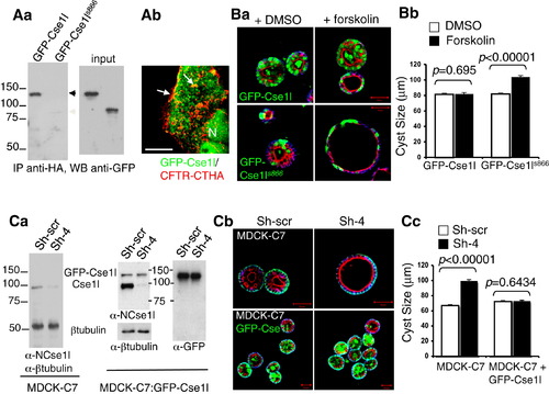
Cse1l Negatively Regulates Fluid Secretion in Mammalian MDCK-C7 Cells (Aa) Zebrafish GFP-Cse1l, but not GFP-Cse1ls866, coimmunoprecipitates with human CFTR-CTHA. (Ab) Partial colocalization (arrows) of GFP-Cse1l and CFTR-CTHA in transfected HEK293 cells. N indicates nucleus. Scale bar represents 10 μm. (B) Overexpression of zebrafish GFP-Cse1l abrogates the stimulatory effect of forskolin on CFTR-dependent fluid secretion in 3D cultures of mammalian MDCK-C7 cells. (Ba) MDCK-C7 cells were grown on Matrigel for 4 days, and forskolin (1 μM) was then added for 16 hr before the cysts were fixed and imaged. Green is GFP, red is F-actin, and blue is β-catenin. Scale bars represent 50 μm. (Bb) Quantification of data from (Ba) (n = 161 for GFP-Cse1ls866; n = 131 for GFP-Cse1l). Error bars indicate standard error of the mean. (C) Depletion of Cse1l leads to increased fluid secretion in 3D cultures of MDCK-C7 cells. (Ca) Immunoblot showing knockdown of dog Cse1l in cells expressing a shRNA targeting Cse1l (Sh-4) but not in control cells expressing a nonsilencing shRNA (Sh-scr). (Cb) Knockdown of dog Cse1l leads to lumen expansion in control but not in GFP-Cse1l-expressing MDCK-C7 cells in 3D cultures (5 days in Matrigel) following forskolin treatment. Red is F-actin, green is β-catenin in upper panels and GFP in lower panels, and blue is ToPro. Scale bars represent 50 μm. (Cc) Quantification of data from (Cb) (n = 200 for control; n = 149 for GFP-Cse1l). Error bars indicate standard error of the mean. |
|
Characterization of baoss866 Mutant Phenotype (Related to Figures 1 and 3) (A) baoss866 mutant enterocytes retain absorptive cell marker expression and localization. 120 hpf WT and baoss866 mutant larvae were fixed in PFA, sectioned and stained for the absorptive marker 4e8. In baoss866 mutant enterocytes, 4e8 (green) was expressed and localized apically as in WT. The arrow points to the apical membrane. Scale bars represent 50 μm. (B) Delaminating cells in baoss866 mutants undergo apoptosis. 120 hpf WT and baoss866 mutant larvae were fixed in PFA, mounted, sectioned and subjected to the Tunel assay to detect apoptotic cells. While all delaminating cells in baoss866 mutant larvae were Tunel positive (red channel, arrows), the proportion of gut cells undergoing apoptosis in WT and baoss866 mutant larvae were essentially the same. Scale bars represent 25 μm. (C) Exocrine pancreas degeneration and liver growth arrest in baoss866 mutant larvae. To visualize endodermal organs, we crossed the baoss866 mutation to a transgenic line expressing GFP and DsRed in the exocrine pancreas and liver, respectively. baoss866 mutant larvae showed a fully penetrant (~100%) exocrine pancreas phenotype (arrows) at 120 hpf. We also detected a variable degree of liver growth arrest (arrowheads) compared to WT siblings. In contrast, we did not detect any obvious pancreatic islet defects (not shown). These phenotypes are fully consistent with cse1l’s expression pattern (see Figure 3F). |
|
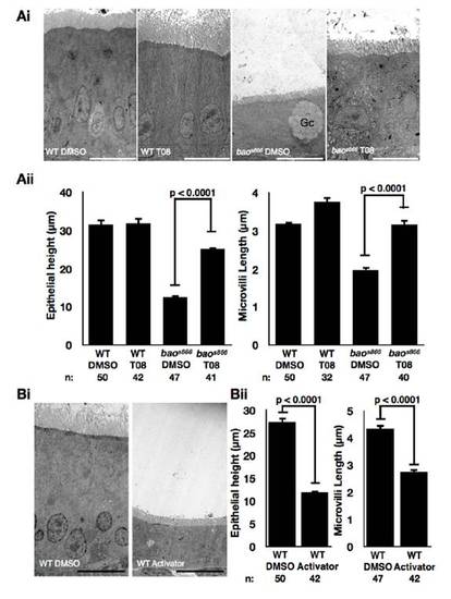
Contribution of CFTR-Dependent Fluid Secretion to Cell Height and Microvilli (mv) Length Reduction in baos866 Mutant Enterocytes (Related to Figure 2) (Ai) TEM micrographs of WT and baos866 mutant larvae treated with DMSO (control) or CFTR inhibitor T08 (5 μM). CFTR inhibition significantly increased cell height (measured from the basal membrane to the base of mv) and mv length in baos866 mutant enterocytes. GC: goblet cell. Scale bars represent 10 μm. (Aii) Quantification of Ai. Error bars: SEM. (Bi) TEM micrographs of WT larvae treated with DMSO (control) or CFTR activator Act9 (15 μM). CFTR activation in WT recapitulated the reduction in cell height and mv length observed in baos866 mutant enterocytes. Scale bars represent 10 μm. PHENOTYPE:
|
|
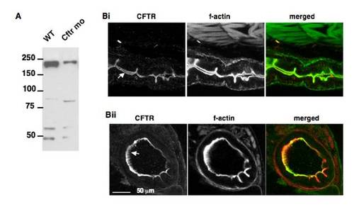
Characterization of Antibodies against Zebrafish CFTR (Related to Figures 2 and 3) (A) In western blots of 120 hpf extracts the antibodies recognized a ~190KDa band. The intensity of the CFTR band was greatly reduced upon anti-sense morpholino knockdown. (Bi) Whole mount staining of 120 hpf larvae showed a tight localization of CFTR to the apical membrane of gut enterocytes (arrow). (Bii) In cross sections of the gut virtually all the CFTR protein was localized to the apical membrane, including the microvilli (arrow). EXPRESSION / LABELING:
|

