
|
Fig. 3 Isolation of the bao Gene
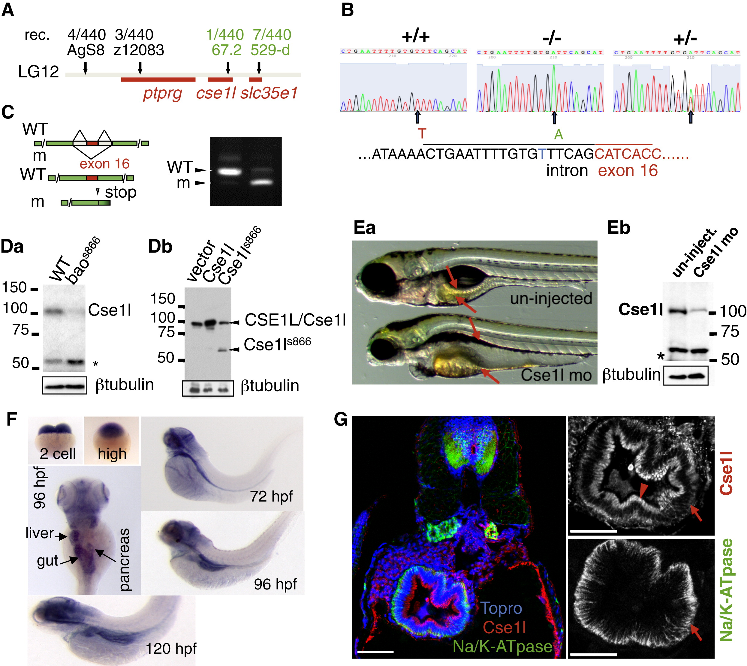
(A) Positional cloning of bao. The number of recombinants (rec.) for each marker is shown.
(B) Sequencing of genomic DNA from +/+, -/-, and +/- larvae revealed a T-to-A mutation (arrow) upstream of the exon 16 splice acceptor site.
(C) Reverse transcriptase PCR on pools of RNA made from WT and baos866 mutants demonstrates defective splicing of exon 16 in baos866 mutants leading to a premature stop codon in exon 17.
(Da) Cse1l is depleted in baos866 mutants. The asterisk marks the position of a cross-reacting protein band.
(Db) The baos866 mutation produces a truncated protein that can be detected in immunoblots of transfected HEK293 cells.
(Ea) Knockdown of Cse1l using an antisense morpholino targeting the translation start site phenocopies the baos866 mutation (22% penetrance at 5 days postfertilization [dpf], n = 220).
(Eb) Immunoblot of 5 dpf uninjected and Cse1l morphants demonstrating knockdown of this protein.
(F) In situ hybridization analysis using an antisense probe directed against the 32 end and untranslated region of the cse1l mRNA.
(G) Immunofluorescence on WT sections showing Cse1l in the subapical (arrowhead) and basolateral (arrow) regions of intestinal cells at 120 hpf. Scale bars represent 50 μm.