
Fig. 2
- ID
- ZDB-FIG-101102-4
- Publication
- Bagnat et al., 2010 - Cse1l Is a Negative Regulator of CFTR-Dependent Fluid Secretion
- Other Figures
- All Figure Page
- Back to All Figure Page
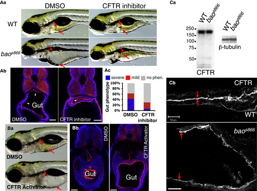
Lumen Expansion in baos866 Mutants Results from Increased CFTR-Dependent Fluid Secretion (A) Inhibition of CFTR blocks lumen expansion in baos866 mutants. (Aa) Bright-field images of WT and baos866 mutants incubated with DMSO (0.1%) or the CFTR inhibitor T08 (5 μM) from 72 to 120 hpf. Arrows point to the edges of the gut lumen. (Ab) Confocal images of cross-sections of 144 hpf control and T08-treated baos866 mutants. Arrowheads point to delaminating cells. Red is F-actin and blue is DAPI. Scale bars represent 50 μm. (Ac) Quantification of the gut phenotype in control and T08-treated baos866 mutants. Larvae were placed in three phenotypic categories (no phenotype, mild phenotype, or severe phenotype) and then genotyped. (B) Activation of CFTR in WT phenocopies the gut lumen expansion defect of baos866 mutants. (Ba) Bright-field image of 144 hpf WT larva treated with DMSO (0.15%) or CFTR-Act9 (15 μM). Arrows point to the edges of the gut lumen. (Bb) Confocal images of cross-sections of 144 hpf control and CFTR-Act9-treated WT larvae. Red is F-actin and blue is DAPI. Scale bars represent 20 μm. (C) Cftr expression and localization is not affected in baos866 mutants compared to WT. (Ca) Immunoblot of 120 hpf WT and baos866 mutants probed against Cftr and β-tubulin. (Cb) Confocal images of whole mounts of 120 hpf WT (top) and baos866 mutant (bottom) stained for Cftr. Arrows point to the apical surface of the gut. Anterior is to the right. Scale bars represent 50 μm. |
| Gene: | |
|---|---|
| Antibody: | |
| Fish: | |
| Anatomical Terms: | |
| Stage: | Day 5 |
| Fish: | |
|---|---|
| Observed In: | |
| Stage: | Day 5 |