- Title
-
Multiple regulatory elements mediating neuronal-specific expression of zebrafish sodium channel gene, Scn8aa
- Authors
- Wu, S.H., Chen, Y.H., Huang, F.L., Chang, C.H., Chang, Y.F., and Tsay, H.J.
- Source
- Full text @ Dev. Dyn.
|
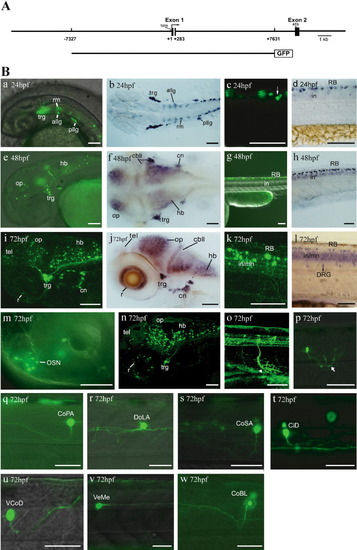
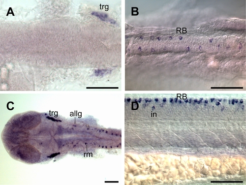
Transient expression profile of scn8aa:GFP. A: The 15-kb fragment of zebrafish scn8aa is cloned into pEGFP-ITR to construct scn8aa:GFP. The gray area in exon 1 (exons are shown in boxes) represents brain-specific regulatory elements shared with mouse SCN8A. B: GFP fluorescence (a, c, e, g, i, k) and in situ hybridization of scn8aa mRNA (b, d, f, h, j, l) in the head and trunk at 24, 48, and 72 hpf. A lateral view is shown, with the anterior to the left and dorsal to the top. m-w: High-magnification views of GFP expression at 72 hpf. Ventral- and dorsal-projecting motoneurons, along with axonal arbors (arrowhead and arrow), are shown in o and p, respectively. GFP-positive interneurons are identified morphologically (q-w). allg, anterior lateral line ganglia; cbll, cerebellum; CiD, circumferential descending interneurons; cn, cranial neurons; CoBL, commissural bifurcating longitudinal interneurons; CoPA, commissural primary ascending interneurons; CoSA, commissural secondary ascending interneurons; DoLA, dorsally longitudinal ascending interneurons; hb, hindbrain; in, interneurons; mn, motoneurons; OP, optic tectum; OSN, olfactory sensory neurons; pllg, posterior lateral line ganglia; r, retina; RB, Rohon-Beard neurons; rm, rhombomere; tel, telecephalon; trg, trigeminal ganglia; VCoD, vental commissural descending interneurons; VeMe, ventral medial interneurons. Scale bars = 100 μm. EXPRESSION / LABELING:
|
|
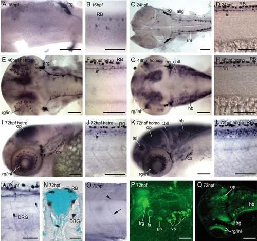
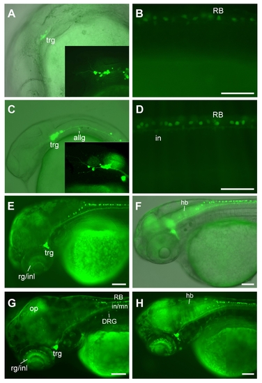
Dorsal and lateral views of GFP expression in Tg(scn8aa:GFP) stable line during embryonic development. A-D: GFP-positive neurons in the head and trunk at 16 and 24 hpf. E-L: GFP-positive neurons in the head and trunk of heterozygotes and homozygotes at 48 hpf (E-H) and 72 hpf (I-L). M-Q: Higher magnification views of specific GFP-expressing neuron types at 72 hpf. M: Lateral view of DRG at the anterior trunk. N: Transverse trunk section of the spinal cord, double-stained with methyl green. The soma of DRG is located outside the spinal cord. O: Axonal projections of ventral and dorsal projecting motoneurons. P: Confocal image of GFP expression in the cranial ganglia and their projections. Q: Confocal image of GFP expression in the optic tectum, trigeminal ganglia (trg), and hindbrain (hb). allg, anterior lateral line ganglia; cbll, cerebellum; cn, cranial neurons; DRG, dorsal root ganglia; fs, facial sensory neurons; gs, glossopharyngeal sensory neurons; in, interneurons; inl, inner nuclear layer; mn, motoneurons; OP, optic tectum; RB, Rohon-Beard neurons; rm, rhombomere; tel, telecephalon; trg, trigeminal ganglia; vs, vagus sensory neurons. Scale bars = 100 μm for A-L and 50 μm for M-Q. EXPRESSION / LABELING:
|
|
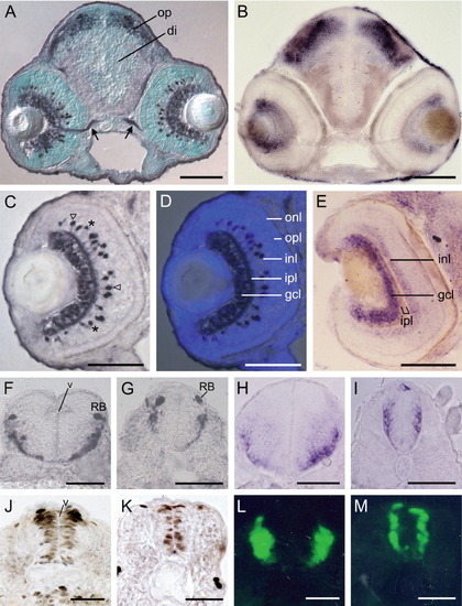
GFP expression in head and trunk regions of Tg(scn8aa:GFP) line at 72 hpf. A: Coronal sections of the head are labeled for GFP antibody and counterstained with methyl green. GFP protein is detected in the retina, optic nerve (arrows), and optic tectum (op), but not in the diencephalon (di). B: Expression pattern of scn8aa mRNA in the brain. C: GFP expression in the retinal ganglia and a subset of interneurons. Open arrowheads show interneurons in the inner nuclear layer with axonal processes. Asterisks show interneurons without axon outgrowth. D: Merged image of the bright field image in C and a corresponding image of DAPI staining. E: Scn8aa mRNA expression in the retinal ganglia cell layer (gcl) and inner nuclear layer (inl). F-I: Cross-sections show GFP expression (F, G) and scn8aa mRNA expression (H, I) in the lateral region of the spinal cord. J, K: BrdU-positive cells at the ventricular zone (v) of the hindbrain and spinal cord. L, M: Early-differentiated neurons of the hindbrain and spinal cord labeled by anti-HuC antibody. ipl, inner plexiform layer; onl, outer nuclear layer; opl, outer plexiform layer; RB, Rohon-Beard neurons. Scale bars = 100 μm for A-E and 50 μm for F-M. EXPRESSION / LABELING:
|
|
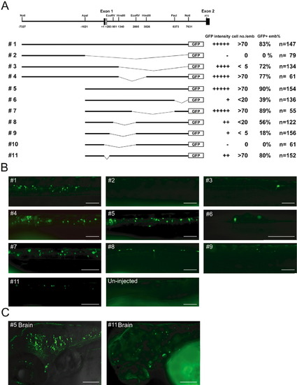
Deletion analysis of the 15-kb regulatory fragment of scn8aa. A: The gray area represents the conserved regulatory element shared with mouse SCN8A. GFP intensity, average number of GFP-positive neurons per embryo (cell no/emb), and percentage of GFP-positive embryos are determined following the transient expression of the indicated deletion construct until 72 hpf. GFP intensity is scored on a six-point scale with “+++++” representing strongest expression and “-” representing no detectable expression. The total number of injected embryos with normal morphology is represented by n. B: Lateral views of GFP expression in embryos injected with the indicated deletion constructs. C: Confocal images of the brains of embryos injected with construct #5 or #11. The #11-injected embryo is subjected to a prolonged exposure due to the low intensity of GFP. Scale bars = 100 μm. |
|
EXPRESSION / LABELING:
|
|
EXPRESSION / LABELING:
|

