
Fig. 2
- ID
- ZDB-FIG-080910-21
- Publication
- Wu et al., 2008 - Multiple regulatory elements mediating neuronal-specific expression of zebrafish sodium channel gene, Scn8aa
- Other Figures
- All Figure Page
- Back to All Figure Page
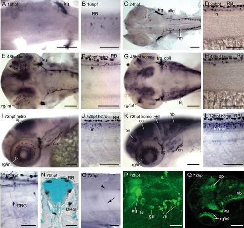
Dorsal and lateral views of GFP expression in Tg(scn8aa:GFP) stable line during embryonic development. A-D: GFP-positive neurons in the head and trunk at 16 and 24 hpf. E-L: GFP-positive neurons in the head and trunk of heterozygotes and homozygotes at 48 hpf (E-H) and 72 hpf (I-L). M-Q: Higher magnification views of specific GFP-expressing neuron types at 72 hpf. M: Lateral view of DRG at the anterior trunk. N: Transverse trunk section of the spinal cord, double-stained with methyl green. The soma of DRG is located outside the spinal cord. O: Axonal projections of ventral and dorsal projecting motoneurons. P: Confocal image of GFP expression in the cranial ganglia and their projections. Q: Confocal image of GFP expression in the optic tectum, trigeminal ganglia (trg), and hindbrain (hb). allg, anterior lateral line ganglia; cbll, cerebellum; cn, cranial neurons; DRG, dorsal root ganglia; fs, facial sensory neurons; gs, glossopharyngeal sensory neurons; in, interneurons; inl, inner nuclear layer; mn, motoneurons; OP, optic tectum; RB, Rohon-Beard neurons; rm, rhombomere; tel, telecephalon; trg, trigeminal ganglia; vs, vagus sensory neurons. Scale bars = 100 μm for A-L and 50 μm for M-Q. |