
- Title
-
Adiponectin and adiponectin receptor genes are coexpressed during zebrafish embryogenesis and regulated by food deprivation
- Authors
- Nishio, S.I., Gibert, Y., Bernard, L., Brunet, F., Triqueneaux, G., and Laudet, V.
- Source
- Full text @ Dev. Dyn.
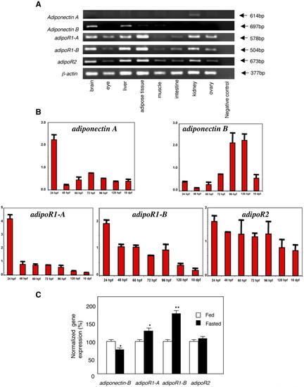
Expression in adult tissues in wild type and fasted fish. A: Semi-quantitative RT-PCR analysis of adiponectin and adipoR genes in various adult tissues. PCR bands were visualized on 1.5% agarose gel stained with ethidium bromide. The PCR amplicons were of expected size, Their sizes are indicated on the right. B: Level of expression of adiponectin and adiponectin receptor genes during zebrafish development. Normalizations of the Q-PCR were done using the 18s gene. Note that all genes are expressed as early as 24 hpf and remain expressed up to 10 dpf. Developmental stages prior 24 hpf were not monitored. C: Q-PCR study of the effects of 96-hr fasting on the expression of the adiponectin and adipoR genes in adult liver. The amount of adiponectin or adipoR mRNA relative to β-actin mRNA is indicated after a normalization at 100% for the fed condition. The data represent mean ± SEM values from three adult zebrafish for each tissue. The stars indicate groups that differ significantly (t-test). |
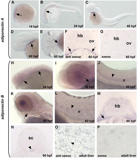
Expression pattern of adiponectin genes during zebrafish embryonic development. Whole mount in situ hybridization of adiponectin A (A-G) and adiponectin B (H-P). A: adiponectin A is first detected at 14 hpf (arrow). B: At 24 hpf, expression expands posteriorly (arrow). C-E: At 48 and 60 hpf, expression is restricted to the anterior part of the pharyngeal region (arrows). F: A transverse section shows that adiponectin A expression is confined to the cell layer in contact with the yolk sac at 60 hpf (arrows). G: A sense probe in a similar section failed to detect any signal. H: adiponectin B is first detected at 24 hpf (arrows). At 36 hpf, mRNA transcripts are detected in the pharyngeal region (arrow) (I) and in the notochord (arrowheads) (J-N). This expression pattern persists until 60 hpf (arrow in K and M; arrowheads in L and N, respectively). O: adiponectin B expression is detected in hepatocytes (arrowhead). P: No signal is detected using the sense probe. sc, spinal cord; hb, hindbrain; n, notochord; ov, otic vesicle. A-D, H-L: Lateral views; E: ventral view; F, G, M-P: transverse sections. EXPRESSION / LABELING:
|
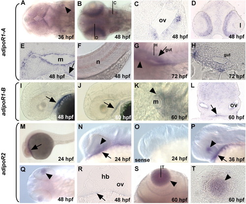
Expression pattern of adipoR genes during zebrafish development. Whole mount in situ hybridization of adipoR1-A (A-H), adipoR1-B (I-L), and adipoR2 (M-T). adipoR1-A is first detected at 36 hpf in the otic vesicle (arrowhead in A) and persists until 48 hpf (B,C). At this developmental stage, adipoR-1A is also detected in the inner nuclear layer of the retina (B, D), around and near the mouth (E), in the pharyngeal region (arrow in E), and in the notochord (F). At 72 hpf, adipoR1-A is present in the liver (arrowhead in G) and in the digestive tract (arrow in G, H). adipoR1-B expression is detected in the pharyngeal region (arrows) at 48 hpf (I) and 60 hpf (J, L). At 60 hpf, adipoR1-B transcripts are also detected in the mouth and in cells near the mouth (arrowhead) (K). adipoR2 is first detected at 24 hpf (arrow in M and N) and in the lens (arrowhead in N). No signal is detected using the adipoR2 sense probe (O). At 36 and 48 hpf, expression remains in the lens and in the pharyngeal region (arrowheads and arrows in P-R). At 60 hpf, expression is solely observed in the lens (S,T). l, lens; m, mouth; n, notochord; ov, otic vesicle. F, G, I, J, M-P: Lateral views; K: ventral view; A, B, Q, S: dorsal views; C, D, E, H, L, R, T: transverse sections. EXPRESSION / LABELING:
|
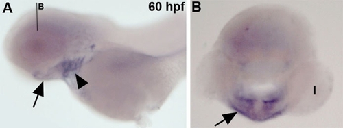
Expression of nkx2.3 at 60 hpf. (A) Lateral view of nkx2.3 expression in the branchial arches (arrowhead) and anteriorly in the ventral pharynx (arrow). (B) Transverse section at the level of the lens (faint line in A) showing that nkx2.3 is expressed as a single continuous cell layer in contact with the yolk (arrowhead). Note that this expression domain is different to what is observed for adiponectin A and B and adipoR2 where two bilateral groups of cells are found instead of a single continuous cell layer. l:lens. EXPRESSION / LABELING:
|