
|
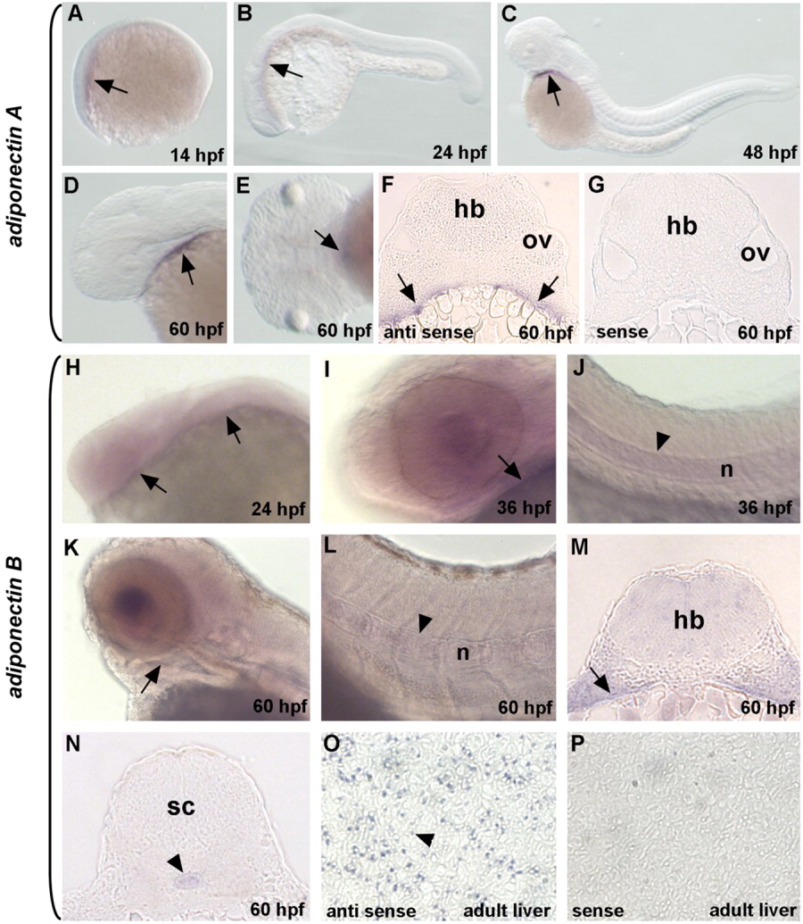
Fig. 3 Expression pattern of adiponectin genes during zebrafish embryonic development. Whole mount in situ hybridization of adiponectin A (A-G) and adiponectin B (H-P). A: adiponectin A is first detected at 14 hpf (arrow). B: At 24 hpf, expression expands posteriorly (arrow). C-E: At 48 and 60 hpf, expression is restricted to the anterior part of the pharyngeal region (arrows). F: A transverse section shows that adiponectin A expression is confined to the cell layer in contact with the yolk sac at 60 hpf (arrows). G: A sense probe in a similar section failed to detect any signal. H: adiponectin B is first detected at 24 hpf (arrows). At 36 hpf, mRNA transcripts are detected in the pharyngeal region (arrow) (I) and in the notochord (arrowheads) (J-N). This expression pattern persists until 60 hpf (arrow in K and M; arrowheads in L and N, respectively). O: adiponectin B expression is detected in hepatocytes (arrowhead). P: No signal is detected using the sense probe. sc, spinal cord; hb, hindbrain; n, notochord; ov, otic vesicle. A-D, H-L: Lateral views; E: ventral view; F, G, M-P: transverse sections.