
- Title
-
Zebrafish brd2a and brd2b are paralogous members of the bromodomain-ET (BET) family of transcriptional coregulators that show structural and expression divergence
- Authors
- Dibenedetto, A.J., Guinto, J.B., Ebert, T.D., Bee, K.J., Schmidt, M.M., and Jackman, T.R.
- Source
- Full text @ BMC Dev. Biol.
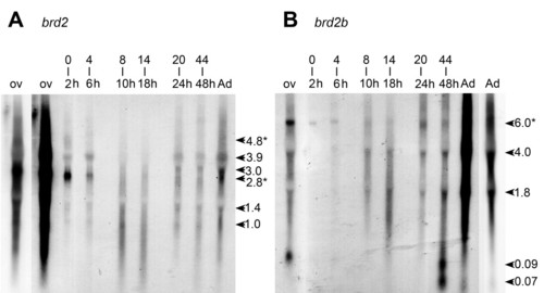
brd2a and brd2b encode gonad-enriched RNAs and multiple developmentally regulated transcripts. Northern blot of total RNA (5 μg/lane) from dissected ovary (ov), adult fish (Ad), and embryos of various stages (0–2, 4–6, 8–10, 14–18, 20–24, and 44–48 hour post fertilization) probed sequentially with A) zf626 (brd2) and B) zf69 (brd2b), radiolabeled cDNA subfragments. RNAs were visualized with EtBr staining and were undegraded and of equivalent amounts in each lane (data not shown). The leftmost ovary lane and the rightmost adult lane are underexposed replicates of adjacent lanes, to allow better band visualization. Ovary-enriched brd2 RNAs (A, 4.8 and 2.8 kb) and ovary-enriched brd2b RNA (B, 6.0 kb) are also present in early embryos (A and B, 0–2 h), and are indicated by asterisks (*). Various size classes of brd2 and brd2b RNAs present in whole adults are also detectable in dissected testis (data not shown). |
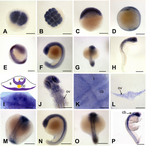
brd2 paralogs are enriched in developing nervous system during segmentation. Whole mount in situ hybridizations to RNA in zebrafish embryos conducted with DIG-labelled zf626 (brd2) (A-L) and zf69 (brd2b) (M-P) cloned sequences. Ubiquitous expression is seen in embryos of 2 cell (A), 16 cell (B), 30% epiboly (C), and bud (D) stages for both brd2 (shown) and brd2b (not shown) genes. Enriched expression in "ladder" patterns along the developing neural keel/rod during segmentation is seen in 14 somite embryos probed for brd2 (E) and for brd2b (M). Cross-sections of 14 somite whole mount embryos probed for brd2 (I, schematic with real image below; 5–6 somite level) show enriched expression in ventrolateral neural rod (nr), floorplate above notochord (nc), and ventral somites (s). Expression is most prominent in head region of 18 somite embyros for both paralogs (F, G; and N, O); only brd2 is also enriched in tail bud (F), while brd2b remains high along the entire ventral trunk (N) at this stage. By 24 hours, brd2 is prominent in the entire head, with some expression in ventral trunk and post-anal tail (H), while brd2b is enriched in ventral brain, cerebellum, and ventral trunk, with low level ubiquitous expression (P). Dorsal flat mounts of 24 hour embryos (J, K) show brd2 expression enriched within caudal tectum (t) at hindbrain-midbrain boundary, within specific cells of the cerebellum (cb) and at the caudal border between cerebellum and rest of hindbrain. The image in (K) is a higher magnification view of a region from another embryo equivalent to the boxed region in J. Expression of brd2 in cells of otic vesicle walls (ov) is apparent both in flat mounts (J) and cross sections at that level (L). Bar = 250 μm for A-H, J, M-P; = 50 μm for I, K; = 100 μm for L. |

brd2 paralogs are differentially expressed in head and trunk regions of pharyngula stage embryos. Whole mount in situ hybridizations to RNA in 44–48 hour zebrafish embryos with DIG-labelled zf626 (A-F) and zf69 (G, H) cloned sequences. Dorsal (A) and lateral (B) views of whole embyros show brd2 expression abundant in all brain subdivisions and developing pharyngeal arches, and in neural retina (nr), otic vesicle (ov), pectoral fin buds (fb) and ventral trunk. Cross sections at levels indicated in A, reveal brd2 expression in: C) mesencephalon (m), diencephalon (d) and neural retina (nr); D) in cells of otic vesicle walls (ov, black arrows); E) in caudal hindbrain (hb), endoderm of prospective digestive tract (en), and pectoral fin bud (fb); F) in developing spinal cord (sc), posterior lateral line primordium (lat), pronephric duct (pn), and endodermal tissue (en), including pancreas progenitor (p) and developing gut (g). Signal in hollow of otic vesicle (white arrows in D) is an artefact due to trapped probe. Although brd2b (G dorsal, H lateral) is also expressed in head region and pectoral fin buds (fb), it is reduced in dorsal midbrain, caudal hindbrain, and ventral trunk, and absent from neural retina (nr, white arrow) and otic vesicle (ov, white arrow). Bar = 250 μm for A,B,G,H; = 100 μm for C, D (two right images); = 25 μm for D (left image). EXPRESSION / LABELING:
|

brd2 paralogs are differentially expressed in head and digestive system of pecfin stage embryos. Whole mount in situ hybridizations to RNA in 60–65 hour zebrafish embryos with DIG-labelled zf626 (A-F) and zf69 (G, H) cloned sequences. Dorsal (A) and lateral (B) views of whole embryos show brd2 expression in all brain subdivisions, especially cerebellum (cb), in neural retina (nr), otic capsule (oc), atrium of heart (h), gill arches (ga), pharynx (ph), swim bladder (sb) and ventral trunk. Cross sections at levels indicated in A reveal brd2 expression in: C) amacrine cells (ac) and ganglion cell layer (gcl) of neural retina; D) spinal cord (sc), pronephric duct (pd) and endodermal derivatives such as liver (li); E) posterior lateral line primordium (lat), and endodermal derivatives such as swim bladder and gut (g). brd2 expression continues in gut (g), but declines in spinal cord (sc, white) as more posterior sections are assayed (F), and is severely reduced in fin bud (D; fb, white) Expression of brd2b (G dorsal, H lateral) is found mainly in head region, but is reduced overall, especially in hindbrain, and nearly absent from otic capsule (oc, white), gill arches (ga, white), heart (h, white), ventral trunk, and endodermal derivatives such as pharanx (ph, white), and swim bladder (sb, white). Bar = 250 μm for A,B,D,G,H; = 100 μm for C,E,F. EXPRESSION / LABELING:
|

brd2 paralogs are maternal transcripts with different patterns of expression in oocytes. Whole mount in situ hybridizations to RNA in zebrafish ovaries with DIG-labelled zf626 (A, C, E, G, I) and zf69 (B, D, F, H, J) cloned sequences. Cross sections through ovaries are shown in E-J. brd2 maternal RNAs are dispersed and abundant in stage I and II oocytes (A, E), and are sometimes found inside the germinal vesicle (gv), as in G (see small oocytes at far right). By stage III,brd2 transcripts accumulate around the outside of the germinal vesicle and rather unevenly around the periphery of the cortex just inside the vitelline envelope (C, E; G, arrowheads). brd2 RNAs then localize around the periphery of the cortex by late stage III, early stage IV (I, arrowheads). brd2 RNAs are also present in follicle cells of stage III/IV oocytes (C; G, I, arrowheads outside oocyte). Although brd2b maternal transcripts are also dispersed throughout stage I and II oocytes, and often found inside the germinal vesicle (B, F, H, J), in contrast to brd2, they become strictly localized to the region surrounding the micropyle (D, mp; H, J, arrowheads), the future animal pole, sometime during stage III, and are not expressed in follicle cells. A,B: 40X; C-F: 80X; G-J, 100X. |
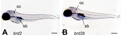
brd2 paralogs are expressed in swim bladder and brain in 5 day old zebrafish. Whole mount in situ hybridizations to RNA in 5 day old zebrafish with DIG-labelled zf626 (A) and zf69 (B) cloned sequences. Lateral views show brd2a and brd2b expression restricted to swim bladder (sb), otic capsule (oc) and brain, with brd2b RNAs most abundant in swim bladder. Bar = 250 μm. EXPRESSION / LABELING:
|

Unillustrated author statements |