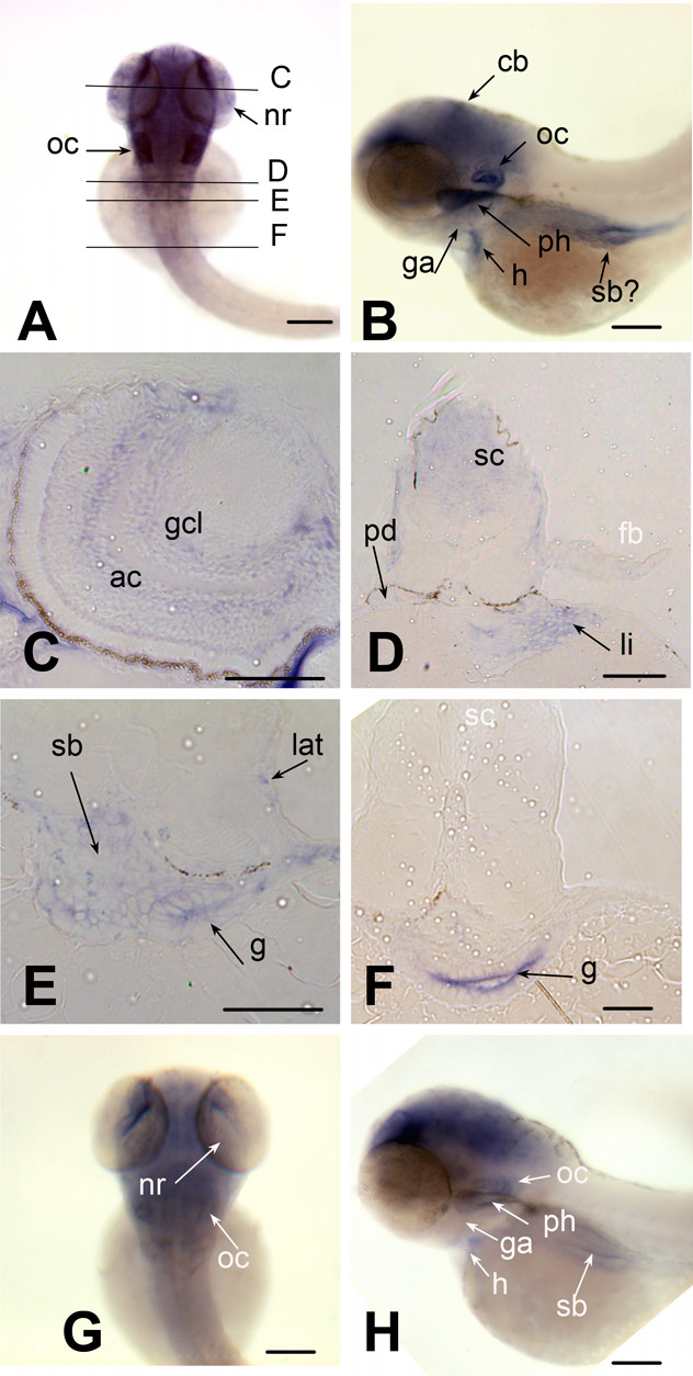
Fig. 7 brd2 paralogs are differentially expressed in head and digestive system of pecfin stage embryos. Whole mount in situ hybridizations to RNA in 60–65 hour zebrafish embryos with DIG-labelled zf626 (A-F) and zf69 (G, H) cloned sequences. Dorsal (A) and lateral (B) views of whole embryos show brd2 expression in all brain subdivisions, especially cerebellum (cb), in neural retina (nr), otic capsule (oc), atrium of heart (h), gill arches (ga), pharynx (ph), swim bladder (sb) and ventral trunk. Cross sections at levels indicated in A reveal brd2 expression in: C) amacrine cells (ac) and ganglion cell layer (gcl) of neural retina; D) spinal cord (sc), pronephric duct (pd) and endodermal derivatives such as liver (li); E) posterior lateral line primordium (lat), and endodermal derivatives such as swim bladder and gut (g). brd2 expression continues in gut (g), but declines in spinal cord (sc, white) as more posterior sections are assayed (F), and is severely reduced in fin bud (D; fb, white) Expression of brd2b (G dorsal, H lateral) is found mainly in head region, but is reduced overall, especially in hindbrain, and nearly absent from otic capsule (oc, white), gill arches (ga, white), heart (h, white), ventral trunk, and endodermal derivatives such as pharanx (ph, white), and swim bladder (sb, white). Bar = 250 μm for A,B,D,G,H; = 100 μm for C,E,F.
Image
Figure Caption
Figure Data
Acknowledgments
This image is the copyrighted work of the attributed author or publisher, and
ZFIN has permission only to display this image to its users.
Additional permissions should be obtained from the applicable author or publisher of the image.
Full text @ BMC Dev. Biol.

