
- Title
-
Zebrafish Angiotensin II Receptor-like 1a (agtrl1a) is expressed in migrating hypoblast, vasculature, and in multiple embryonic epithelia
- Authors
- Tucker, B., Hepperle, C., Kortschak, D., Rainbird, B., Wells, S., Oates, A.C., and Lardelli, M.
- Source
- Full text @ Gene Expr. Patterns
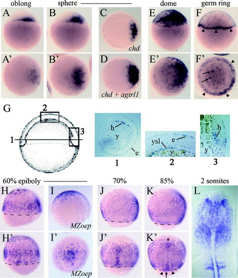
Expression of agtrl1a during blastula and gastrula stages In situ transcript hybridisations on embryos up to 11 hpf. (A–F) Expression pattern of agtrl1a through blastula stages. Lateral views A, B, E, and F (upper panels) and animal pole views A′, B′, E′, F′ (lower panels), all with dorsal to right. C shows dorsal expression domain of chd, D shows embryo co-hybridized with chd and agtrl1a riboprobes. In F and F′ arrowheads marks the germ-ring and arrows indicate the YSL nuclei. (G) Sections through 6 hpf embryos after in situ transcript hybridisation against agtrl1a. The diagram shows the positions of the sections. (1) Horizontal section through mesendodermal part of the embryo (shield to top), (2) vertical section through the embryo, (3) vertical section through the shield; y, yolk; e, enveloping layer, h, hypoblast, ysl, yolk syncitial layer. (H–K) Expression of agtrl1a during gastrula stages. Lateral views with dorsal to right H–K, dorsal views H′, J′, K′ and animal view I′. Dashed lines mark the gastrula margin; in K′ the asterisk, arrow and the arrowheads mark the prechordal plate, the axial mesoderm, and the adaxial cells, respectively. I, I′ shows agtrl1a expression in MZoep embryos. (L) Flat mounted embryo with anterior up, showing agtrl1a expression in the head. EXPRESSION / LABELING:
|
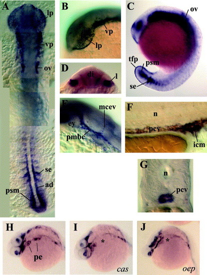
Expression of agtrl1a during segmentation and pharyngula stages in situ transcript hybridisations showing agtrl1a expression in embryos from 14 to 24 hpf. (A) Dorsal axial view of 14 hpf embryo. Expression is observed in the epithelium covering the retina (lens primordium, lp), putative vascular precursors (vp) lateral to mid- and hindbrain, in otic vesicles (ov), in somitic epithelia (se), presomitic mesoderm (psm) and adaxial cells (ad). (B) Lateral view of head of embryo in A. (C) Lateral view of an embryo at 18.5 hpf showing agtrl1a expression in otic vesicles (ov), tail fin primordium (tfp), presomitic mesoderm (psm) and the most recently formed somite epithelium (se). (D) Transverse section at the level of the diencephalon (di) at 24 hpf showing expression in the developing lens (l). Dorsal is up. (E) Lateral view of the developing head of an embryo at 24 hpf showing agtrl1a expression in developing vasculature. The primordial midbrain channel (pmbc), middle cerebral vein (mcev) and developing eye (ey) are indicated. (F) Lateral view of the yolk extension and cloaca region of an embryo at 24 hpf showing expression in the primary caudal vein (pcv) and intermediate cell mass (icm). (G) Transverse section at the level of the yolk extension at 24 hpf showing expression in the primary caudal vein. (H–J) agtrl1a expression in the pharyngeal endoderm (pe) in wt (H), is absent in cas (I) and oep (J) embryos (asterisk) at 24 hpf seen in lateral oblique view, anterior to left. EXPRESSION / LABELING:
|
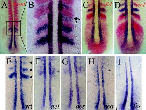
Expression of agtrl1a in somites and in mutants affecting somitogenesis (A–D) In situ transcript hybridisations of 15–16 hpf (12–14 somite) wild-type embryos, dorsal views over the posterior notochord (centre), anterior to top. (A and B) agtrl1a and myod; (C) agtrl1a and dld; (D) agtrl1a and her1: agtrl1a expression is stained blue while myod, her1 and dld expression is red. B is a magnification of the region marked with the box in A; a, anterior somite half, p, posterior somite half, arrow indicates the most recently formed somite furrow. (E–I) In situ transcript hybridisation to detect agtrl1a expression at 16 hpf in mutant zebrafish embryos. (E) Wild-type embryo, normal somites are indicated with arrowheads. Embryos homozygous for the mutations (F) after eight (aeitr233), (G) deadly seven (destp37), (H) beamter (beatm98) with region of disrupted agtrl1a expression indicated with an asterisk, and (I) fused somites/tbx24 (fss/tbx24te314a). EXPRESSION / LABELING:
|
Reprinted from Gene expression patterns : GEP, 7(3), Tucker, B., Hepperle, C., Kortschak, D., Rainbird, B., Wells, S., Oates, A.C., and Lardelli, M., Zebrafish Angiotensin II Receptor-like 1a (agtrl1a) is expressed in migrating hypoblast, vasculature, and in multiple embryonic epithelia, 258-265, Copyright (2007) with permission from Elsevier. Full text @ Gene Expr. Patterns