
- Title
-
Lithium perturbation and goosecoid expression identify a dorsal specification pathway in the pregastrula zebrafish
- Authors
- Stachel, S.E., Grunwald, D.J., and Myers, P.Z.
- Source
- Full text @ Development
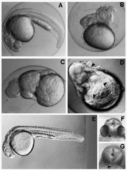
LiCl produces three classes of defects in zebrafish embryos. Embryos were exposed to 0.3 M LiCl for 10 minutes at the 64-cell stage (2 h; B-D), and at the sphere stage (4 h; E and G), and photographed at 26 h (AD) or 28 h (E-F). (A) Control embryo. (B) Bustled embryo. (C) Radialized embryo. (D) Radialized embryo. Arrowheads indicate distinct notochord-like structures. (E) Eyedefective embryo. (F) Dorsoanterior view of a control embryo CNS, with the first (*) and third (arrowhead) ventricles indicated. (G) Equivalent view of an anophthalmic embryo. |
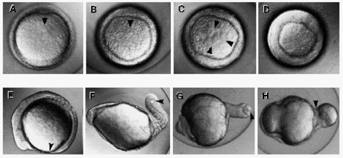
Early lithium exposure leads to excess shield formation and aberrant morphogenesis. (A-D) Animal pole views of 6 h embryos. Arrowheads mark the location and presumptive direction of migration of regions of the germ ring having embryonic shield morphology. (A) Control embryo. The shield appears as an anterior thickening of the germ ring and occupies a 30° arc. (B) Lithiumized embryo, broadened shield. A single thickening at the germ ring subtends a 90° arc. (C) Lithiumized embryo, supernumerary shields. (D) Lithiumized embryo, radialized shield. (E-H) Lateral views of 12.5 h embryos. Anterior is to the left in each panel. The blastopore in each embryo is indicated by an arrowhead. (E) Control embryo. Seven somites have formed, and the tailbud, which is just caudal of the blastopore, has not yet extended off the yolk. (F) Lithiumized embryo. A large mass of segmented tissue extends posteriorly, with the blastopore located at its terminus. (G) Lithiumized embryo. The large posterior extension is unsegmented. (H) Lithiumized embryo. The blastopore has closed onto and bisected the yolk, and the extruded portion of the yolk is partially covered by posteriorly extending cellular material. |
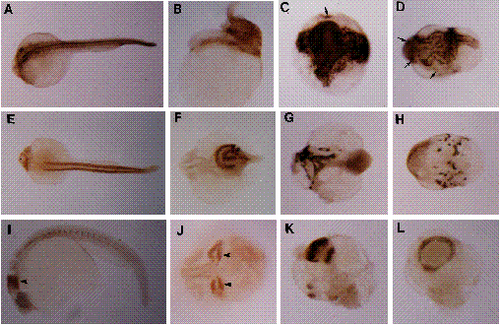
Immunohistochemical analysis of hyper-dorsal development in 26-28 h lithiumized embryos. Anterior is to the left in all panels for which orientation can be determined. (A-D) Analysis of notochord differentiation with MZ15. (A) Control embryo, dorsal view. The anterior end of the notochord sheath is just behind the ears. (B) Bustled embryo, lateral view. The notochord is highly twisted and its anterior end is much more posteriorwards than for the control. (C) Radialized embryo, vegetal-pole view. Several distinct and twisted notochords radiate anteriorwards from a large mass of stained tissue that surrounds the blastopore, which has prematurely closed onto the yolk. (D) Radialized embryo, indeterminate orientation. Arrows in C and D indicate distinct patches of notochord tissue. (E-H) Analysis of skeletal muscle differentiation with F59. (E) Control embryo, dorsal view. Heart staining is to the left, anterior to the ears. The somite muscle appears as two lateral bands down the length of the trunk. (F) Bustled embryo, dorsal view. No heart staining is seen. The skeletal staining appears as two twisted lateral bands. (G) Radialized embryo, indeterminate orientation. The muscle staining, which is greatly reduced in relative amount, is highly disorganized. (H) Radialized embryo, indeterminate orientation. Several small patches of muscle tissue occur around the circumference of this specimen. (I-L) Analysis of neural engrailed expression with 4D9. (I) Control embryo, lateral view. Neural engrailed staining describes a band of cells at the midbrain-hindbrain junction, and muscle engrailed staining marks muscle pioneer cells located bilaterally within each somite. (J) Bustled embryo, dorsal view. A single anterior axis bifurcates at the level of the diencephalon to produce two foci of neural engrailed expression. Arrows in I and J indicate neural staining. (K) Radialized embryo, indeterminate orientation. Multiple sites of neural engrailed staining are observed. (L) Radialized embryo. Neural staining describes a complete band around the embryo, indicating that neural induction has occurred radially versus linearly. Such staining can coincide with the position of the blastopore in embryos that fail to complete epiboly. Note that radialized engrailed staining is similarly observed in lithiumized Xenopus embryos (Hemmati-Brivanlou and Harland, 1989). |
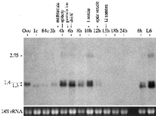
Northern blot analysis of goosecoid expression in oocytes, staged embryos and LiCl-treated embryos. 4 μg samples of total RNA from zebrafish oocytes (mixed stages) and embryos of the indicated stages were fractionated on a 1% formaldehyde-agarose gel, transferred to a nylon membrane and probed with the entire 1.3 kb pZG10.3 cDNA insert. RNA concentrations were estimated from A260 measurements, and the 18S rRNA band intensities are shown at the bottom. Note that roughly three-fold less RNA was loaded in the 8 hour versus the adjacent lanes. Developmental stages correspond to hours post-fertilization except Ooc, 1c, 64c, and L6, which respectively correspond to oocytes (mixed stages), 1-cell embryos (15-30 minutes postfertilization), 64-cell embryos (2 hours postfertilization) and 6 h lithiumized embryos. Developmental landmarks and the sizes of the three major goosecoid transcripts are indicated at the top and left of the figure. Transcript sizes were calculated from the mobilities of RNA standards. |
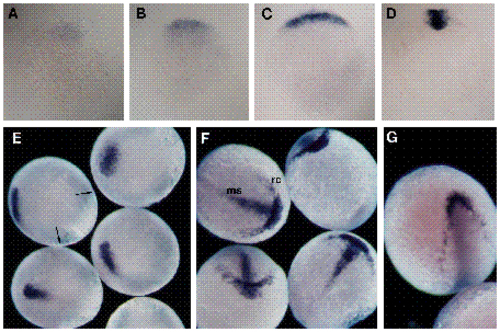
Spatial localization of goosecoid transcripts during embryogenesis. Developmental times are +/- 15 minutes. The blue staining corresponds to goosecoid hybridization. For A-D, the yolks were removed and the blastoderms were photographed in bright field. (A) Animal pole view of ‘early’ 4 h embryo. Expression is limited to a faint patch of cells. (B) Animal pole view of ‘late’ 4 h embryo. The expression domain has expanded to describe a 90° sector. (C) Animal pole view of 5 h embryo with staining limited to cells along the margin. Most cells within the expression domain exhibit staining by this stage. (D) Vegetal pole view of 6 h embryo. Hybridization is restricted to involuted cells within the shield. For E-G, the yolks were left intact, and the embryos were photographed in dark field. (E) Lateral and dorsal views of 8 h embryos. The arrows in the upper left embryo indicate the blastopore at the germ-ring/yolk margin. (F) Anterior and dorsal views of 10 h embryos. The blastopore has closed just ventral to the vegetal pole and the anterior edge of the hybridization is at the animal pole. The medial-strip (ms) and rostral crescent (rc) patterns are indicated in the upper left embryo. The rostral crescent is seen to occupy a position superficial to that of the medial-strip in the top embryo. Staining is sporadic in the lateral regions of the wing-like extensions of the rostral crescent. (G) Dorsal view of 12 h embryo, with sporadic staining along its anterolateral margins. Medial staining is no longer seen. EXPRESSION / LABELING:
|
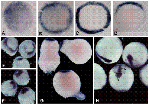
Spatial localization of goosecoid transcripts in lithiumized embryos. Stages are +/- 15 minutes. Embryos in A-G and in H were exposed to 0.3 M LiCl for 10 minutes at 2 h and 4 h, respectively. For A-D, the yolks were removed and the blastoderms were photographed in bright field. (A) Animal pole view of a 4 h embryo. Staining is observed throughout the blastoderm; this staining is typically slightly more intense in a portion of the blastoderm. (B) Animal pole view of a 5 h embryo. While significant hybridization still occurs throughout the blastoderm, the staining is largely concentrated around the margin. (C) Animal pole view of 6 h embryo, radialized shield. Cells around the entire circumference of the germ ring are stained. (D) Animal pole view of a 6 h embryo, broad shield. Staining is restricted to a 160° arc at the margin. Note that lateral observation of the 6 h embryos shows that staining is restricted to involuted cells. For E-H, the yolks were left intact, and the embryos were photographed in dark field. (E) Oblique animal pole view of 8 h embryos. A ring of hybridization is observed above the germ-ring/yolk margin. (F) Oblique animal pole view of 10 h embryos. Hybridization is just ventral to the animal pole. (G) Lateral view of 14 h embryos. A circular cap of staining occupies the animal pole. (H) Dorsal and anterior views of 10 h late-lithium embryos. While the medial-strip (ms) and medial swelling of the rostral-crescent (rc) patterns are observed, the lateral-wing staining is reduced or absent. EXPRESSION / LABELING:
|

Unillustrated author statements EXPRESSION / LABELING:
|