
Fig. 4
- ID
- ZDB-FIG-060113-16
- Publication
- Stachel et al., 1993 - Lithium perturbation and goosecoid expression identify a dorsal specification pathway in the pregastrula zebrafish
- Other Figures
- All Figure Page
- Back to All Figure Page
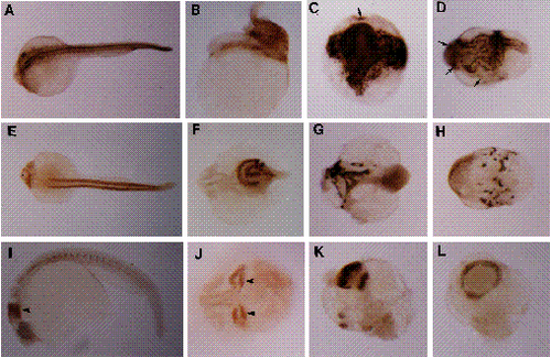
Immunohistochemical analysis of hyper-dorsal development in 26-28 h lithiumized embryos. Anterior is to the left in all panels for which orientation can be determined. (A-D) Analysis of notochord differentiation with MZ15. (A) Control embryo, dorsal view. The anterior end of the notochord sheath is just behind the ears. (B) Bustled embryo, lateral view. The notochord is highly twisted and its anterior end is much more posteriorwards than for the control. (C) Radialized embryo, vegetal-pole view. Several distinct and twisted notochords radiate anteriorwards from a large mass of stained tissue that surrounds the blastopore, which has prematurely closed onto the yolk. (D) Radialized embryo, indeterminate orientation. Arrows in C and D indicate distinct patches of notochord tissue. (E-H) Analysis of skeletal muscle differentiation with F59. (E) Control embryo, dorsal view. Heart staining is to the left, anterior to the ears. The somite muscle appears as two lateral bands down the length of the trunk. (F) Bustled embryo, dorsal view. No heart staining is seen. The skeletal staining appears as two twisted lateral bands. (G) Radialized embryo, indeterminate orientation. The muscle staining, which is greatly reduced in relative amount, is highly disorganized. (H) Radialized embryo, indeterminate orientation. Several small patches of muscle tissue occur around the circumference of this specimen. (I-L) Analysis of neural engrailed expression with 4D9. (I) Control embryo, lateral view. Neural engrailed staining describes a band of cells at the midbrain-hindbrain junction, and muscle engrailed staining marks muscle pioneer cells located bilaterally within each somite. (J) Bustled embryo, dorsal view. A single anterior axis bifurcates at the level of the diencephalon to produce two foci of neural engrailed expression. Arrows in I and J indicate neural staining. (K) Radialized embryo, indeterminate orientation. Multiple sites of neural engrailed staining are observed. (L) Radialized embryo. Neural staining describes a complete band around the embryo, indicating that neural induction has occurred radially versus linearly. Such staining can coincide with the position of the blastopore in embryos that fail to complete epiboly. Note that radialized engrailed staining is similarly observed in lithiumized Xenopus embryos (Hemmati-Brivanlou and Harland, 1989). |