- Title
-
Zebrafish etv2 knock-in line labels vascular endothelial and blood progenitor cells
- Authors
- Chestnut, B., Sumanas, S.
- Source
- Full text @ Dev. Dyn.
|
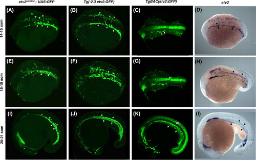
A comparison of etv2 ci32Gt+/?; UAS:GFP, Tg(?2.3etv2:GFP), TgBAC(etv2:GFP) fluorescence pattern and etv2 mRNA expression analyzed by in situ hybridization (ISH) at the 14?21 somite stages. A?C, GFP fluorescence or, D, etv2 mRNA expression is apparent in bilaterally located vascular endothelial progenitors which have started migrating toward the midline at the 14?15?somite stages (arrowheads). Strong nonspecific GFP expression in the neural tube (NT) is apparent in TgBAC(etv2:GFP) embryos. E?H, GFP?positive vascular endothelial progenitors at the 18?19?somite stages are coalescing at the midline into vascular cords (arrowheads, E,F). Similar etv2 expression pattern is observed by ISH analysis, H. I?L, GFP expression at the 20?21?somite stages is apparent in the forming axial vasculature (arrowheads), and nonspecific expression is apparent in the neural tube, K. Similar etv2 expression pattern is apparent from ISH analysis, L. A?H, dorsal or dorsolateral view; I?L, lateral view, anterior is to the left EXPRESSION / LABELING:
|
|
A comparison of etv2 ci32Gt+/?; UAS:GFP, TgBAC(etv2:GFP), Tg(?2.3etv2:GFP) fluorescence pattern (in kdrl:mCherry background, top row) and etv2 mRNA expression analyzed by in situ hybridization (ISH) at 24 hpf. etv2 ci32Gt+/?; UAS:GFP expression is apparent throughout the entire vasculature, red blood cells (RBC), and macrophages (MF). TgBAC(etv2:GFP) shows similar expression pattern, and also shows nonspecific expression in the neural tube. A?C, Merged mCherry and GFP channels; D?F, GFP channel; G,H, magnified images of the trunk region in, A and B. I, ISH analysis for etv2 mRNA expression at 24 hpf. DA, dorsal aorta; PCV, posterior cardinal vein; ISV, intersegmental vessels EXPRESSION / LABELING:
|
|
A comparison of etv2 ci32Gt+/?; UAS:GFP, TgBAC(etv2:GFP), Tg(?2.3etv2:GFP) fluorescence pattern (in kdrl:mCherry background) at 48 hpf, A?I, and 72 hpf, J?R. etv2 ci32Gt+/?; UAS:GFP expression is apparent throughout the entire vasculature and in lymphatic progenitors (parachordal lymphangioblasts, PLs). TgBAC(etv2:GFP) shows similar expression pattern, and also shows nonspecific expression in the neural tube. Vascular endothelial expression in Tg(?2.3etv2:GFP) line is downregulated after 48 hpf and is very weak at 72 hpf, while nonspecific epithelial expression is apparent. A?C, J?L, Merged mCherry and GFP channels; D?F, M?O, GFP channel; G?I, P?R, magnified images of the trunk region in, A?C, J?L. DA, dorsal aorta; PCV, posterior cardinal vein; ISV, intersegmental vessels, SIV, subintestinal vessel; DLAV, dorsal longitudinal anastomotic vessel; CCV, common cardinal vein EXPRESSION / LABELING:
|
|
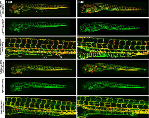
A comparison of etv2 ci32Gt+/?; UAS:GFP and TgBAC(etv2:GFP) embryos in kdrl:mCherry background at 5 and 7 dpf. Both lines show GFP expression in the entire vasculature and lymphatics. DA, dorsal aorta; PCV, posterior cardinal vein; SIV, subintestinal vein (thoracic duct, TD). Tg (?2.3etv2:GFP) line did not show vascular endothelial expression at these stages |
|
etv2 expression analysis in wild?type, etv2 ci32G?+??/? and etv2 ci32Gt?/? embryos at the 20?somite, A?C, and 24 hpf stages, D?F. Embryos were obtained from an incross of etv2 ci32Gt+/?; UAS:GFP +/+ parents and sorted based on their GFP fluorescence pattern. Antisense etv2 RNA probe which corresponds to the C?terminal portion of the etv2 coding sequence and 3?UTR downstream of the Gal4 insertion side was used for in situ hybridization (see Experimental Procedures). A?C, In non?fluorescent wild?type siblings (wt), strong etv2 expression is apparent in vascular progenitors in the anterior lateral plate mesoderm, presumptive progenitors of the anterior and common cardinal veins, and in vascular progenitors next to the tailbud (arrows). Weaker expression in the dorsal aorta (DA) is also apparent. Note the reduced etv2 expression in etv2 ci32Gt+/? embryos and nearly absent expression in etv2 ci32Gt?/? embryos. D?F, In wild?type siblings, strong etv2 expression is apparent in the cranial venous vasculature, including the primordial midbrain channel (PMBC) and the middle cerebral vein (MCEV), as well as the tail plexus region (arrow). Weaker expression in the DA is also apparent. Note the reduced etv2 expression in etv2 ci32Gt+/? embryos and nearly absent expression in etv2 ci32Gt?/? embryos EXPRESSION / LABELING:
PHENOTYPE:
|
|
A comparison of vascular development in heterozygous etv2 ci32Gt+/?; UAS:GFP and homozygous etv2 ci32Gt?/?; UAS:GFP embryos. GFP expression is apparent in vascular progenitors of the trunk axial vasculature (arrow, A) in etv2 ci32Gt+/?; UAS:GFP embryos. This domain is broader and disorganized in etv2 ci32Gt?/?; UAS:GFP embryos, E. Vascular progenitors fail to coalesce into vascular cords in etv2 ci32Gt?/?; UAS:GFP embryos at 25 hpf (arrows, F, compare with C). Intersegmental vessels (ISVs) are absent in etv2 ci32Gt?/?; UAS:GFP embryos between 25 and 48 hpf, F and G. Some ISV sprouts are apparent at 72 hpf but are shorter and mispatterned (H, arrows) EXPRESSION / LABELING:
PHENOTYPE:
|
|
Brightfield and fluorescent images of etv2 ci32Gt+/? and etv2 ci32Gt?/? embryos and their non?fluorescent wild?type siblings at 24 hpf. No morphological defects are apparent in the brightfield images of etv2 ci32Gt+/? and etv2 ci32Gt?/? embryos PHENOTYPE:
|


