- Title
-
Transgenic retinoic acid sensor lines in zebrafish indicate regions of available embryonic retinoic acid
- Authors
- Mandal, A., Rydeen, A., Anderson, J., Sorrell, M.R., Zygmunt, T., Torres-Vazquez, J., and Waxman, J.S.
- Source
- Full text @ Dev. Dyn.
|
RA sensor reporters are expressed in established domains of RA responsive gene expression. A, B, K, L: Reporter expression in both the Tg(β-actin:GDBD-RLBD);(UAS:EGFP) and Tg(β-actin:GDBD-RLBD);(UAS:mCherry) lines initiates in the spinal cord (arrows in A and K) by the tailbud (TB) stage. C, D, M, N: RA sensor reporter expression is strengthened in the anterior spinal chord by the 8-somite (s) stage (arrows in C and M). E, F, O, P: RA sensor reporter expression is maintained in the anterior spinal cord (sc) and noticeable expanded to the adjacent tissues, including the notochord (nc) and ectoderm (ec), by the 16s stage. G, H, Q, R: RA sensor reporter expression was maintained in the tissues of the mid-region (anterior trunk and spinal cord), but was now more easily observed in the somites (sm) and pronephros (pn). I, J, S, T: Both the Tg(β-actin:GDBD-RLBD);(UAS:EGFP) and Tg(β-actin:GDBD-RLBD);(UAS:mCherry) lines initiate some expression in the brain by 36 hpf. However, we only found eye expression (arrow in I) in the Tg(β-actin:GDBD-RLBD);(UAS:EGFP) line. Views in A, C, E, G, I, K, M, O, Q, S are lateral. Views in B, D, F, H, J, L, N, P, R, T are dorsal. In all images, anterior is up. |
|
Fluorescence in the Tg(β-actin:GDBD-RLBD);(UAS:EGFP) and Tg(β-actin:GDBD-RLBD);(UAS:mCherry) lines. A, E: Fluorescence in the Tg(β-actin:GDBD-RLBD);(UAS:EGFP) line is found in the spinal cord (arrow) by the 16s stage, although expression in the Tg(β-actin:GDBD-RLBD);(UAS:mCherry) is typically not visible by this stage. B, F: Expression at 24 hpf is visible in the spinal cord, notochord, and somites. Expression in the adjacent non-spinal cord tissues is more easily visible in the Tg(β-actin:GDBD-RLBD);(UAS:EGFP) embryos. C, D, G, H: Expression in maintained in the spinal cord, notochord, and somites at 30 and 48 hpf. Expression is seen in the ventral eye by 48 hpf (arrow in D). Arrowhead in D indicates the somites. Expression in the eye of the Tg(β-actin:GDBD-RLBD);(UAS:mCherry) embryo is from the α-cry:DsRed used in the transgenic construct. Arrow in H indicates autofluorescent pigment cells. All views are lateral with anterior up. |
|
RA sensor lines are responsive to RAR agonists and antagonists. A, A2, H, H2: Control untreated embryos. B, B2, C, C2, I, I2, J, J2: RA- or AM580-treated embryos show almost ubiquitous expression of the reporter. Embryos treated with RA or AM580 were posteriorized as a consequence of treatment with these agents. D, D2, K, K2: Keto caused an expansion of reporter expression. E, E2, F, F2, L, L2, M, M2: DEAB or RO41 treatment inhibited expression. None of the DEAB-treated (0 of 17) or R041-treated (0 of 17) Tg(β-actin:GDBD-RLBD);(UAS:EGFP) embryos had expression compared to 28% (17 of 61) sibling embryos, which is close to the 25% of expected fluorescent embryos used from an intercross of Tg(β-actin:GDBD-RLBD) and Tg(UAS:EGFP) hemizygous individuals. None of DEAB-treated (0 of 15) or R041-treated (0 of 14) Tg(β-actin:GDBD-RLBD);(UAS:mCherry) embryos had expression compared to 34% (18 of 53) sibling embryos, which is close to the 37.5% of expected fluorescent embryos used from an intercross of Tg(β-actin:GDBD-RLBD);(UAS:mCherry) hemizygous and (UAS:mCherry) hemizygous individuals. A–F and H–M are fluorescent images of live embryos. A2–F2 and H2–M2 are ISH. Arrows in A,A2,D,D2,H,H2,K,K2 indicate the length of the domain of reporter expression. All views are lateral with anterior up. |
|
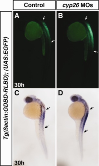
RA sensor expression is expanded in Cyp26-deficient embryos. A, C: Control uninjected sibling embryos. B, D: The domain of reporter expression is expanded in embryos injected with MOs targeting cyp26a1 and cyp26c1. Arrows indicate the length of expression. Views are lateral with anterior up. |
|
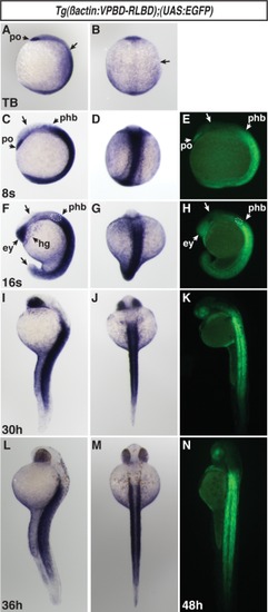
Expression in Tg(β-actin:VPBD-RLBD);(UAS:EGFP) embryos is consistent with the domains of RA-producing and -degrading enzymes. A, B: VP16-RA sensor expression at the TB stage. Arrowhead indicates strong expression in the polster (po). Arrows in A and B indicate the border between the posterior reporter expression and the anterior of the embryo, which lacks reporter expression. C–E: VP16-RA sensor expression via ISH and fluorescence at the 8s stage. Expression is maintained in the polster (po) and is largely absent in the anterior brain (arrows in C and E). There is strong expression throughout the posterior. Lower levels of expression extend into the posterior hindbrain (phb). F–H: VP16-RA sensor expression via ISH and fluorescence at the 16s stage. There is expression in the eyes (ey) and anterior brain, hatching gland (hg), and posterior hindbrain. Expression extends into the posterior hindbrain up to the otic vesicle (dashed outlines in F and H), which forms at the rhombomeres 5 and 6. Expression is absent in the brain and tip of the tail (arrows in F and H). I–K: RA reporter expression via ISH and fluorescence at 30 hpf. L, M: VP16-RA sensor expression via ISH at 36 hpf. N: VP16-RA sensor expression via fluorescence at 48 hpf. A, D, F, G, I, J, L, M, O are lateral views. B, E, H, K, N are dorsal views. In all images anterior is up. |
|
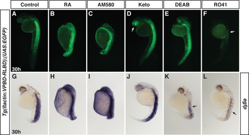
Tg(β-actin:VPBD-RLBD);(UAS:EGFP) embryos are sensitive to RAR agonists and antagonists. A, G: Control sibling embryos at 30 hpf. B, C, H, I: RA- and AM580-treated VP16-RA sensor embryos have almost ubiquitous expression. Embryos are posteriorized due to RAR agonist treatment. D, J: VP16-RA sensor embryos treated with Keto have expanded and enhanced expression, particularly in the anterior brain and eye (arrow in D). E, F, K, L: VP16-RA sensor embryos treated with DEAB or RO41. By ISH, DEAB and RO41 reduced expression (arrows in K and L). However, RO41 was more effective. By fluorescence, it was often hard to distinguish a loss in reporter expression in DEAB-treated embryos. While we observed reductions in reporter expression, particularly with RO41 treatment, we never completely lost expression in embryos treated with RAR or Aldh1a antagonists. All views are lateral with anterior up. |
|
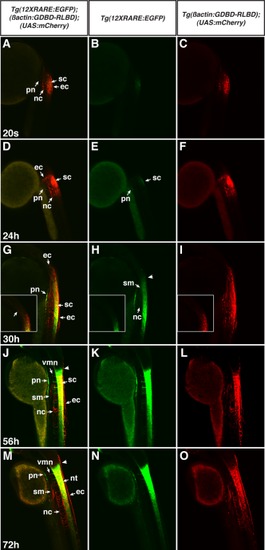
Direct comparison of the 12XRARE:EGFP and β-actin:GDBD-RLBD;UAS:mCherry transgenes. A–F: At the 20s stage and 24 hpf, the RA sensor is expressed in the skin epithelial cells (ec), the spinal cord (sc), the pronephros (pn) and the notochord (nc), while the 12XRARE:EGFP transgene is less strongly visible in the spinal cord and pronephros. G–I: By 30 hpf, mCherry expression from the β-actin:GDBD-RLBD;UAS:mCherry reporter has expanded in the anterior-posterior axis, but the tissue types expressing the reporter have not changed. MCherry can be seen in axons projecting anteriorly from the spinal cord (G). Insets in G–I are parallel confocal slices to the images in G–I, but focus on the anterior border of expression from the reporters. 12XRARE:EGFP reporter expression has also expanded in the A–P axis and the intensity of the reporter has increased. Low levels of expression are also in the somites (sm), though this is more obvious from mCherry. J–L: By 56 hpf, expression of both reporters has expanded more posteriorly, while the anterior border of expression at the hindbrain–spinal cord boundary has become more distinct. Expression is also seen in the axonal projects from the ventral motor neurons (vmn). M–O: Expression in these same tissues is maintained past 72 hpf. The ventral motor neurons can be seen elaborating further. All images are single confocal slices. Embryos are lateral views with anterior up. |
|
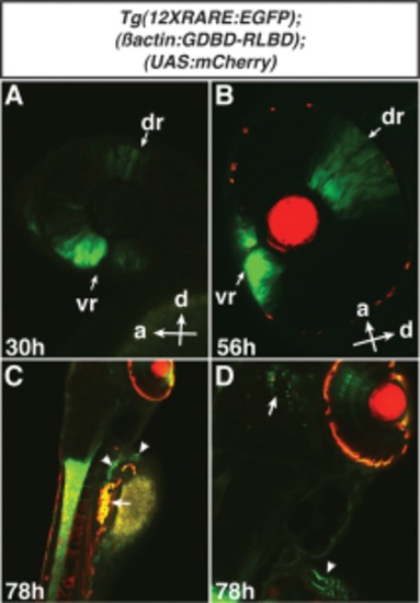
The 12XRARE:EGFP transgene is expressed in the eye, pectoral fin motor neurons, and brain. A: The 12XRARE:EGFP reporter is expressed in the dorsal retina (dr), by 30 hpf. The dorsal retina expression was visible due to PTU treatment. Expression in the ventral retina (vr) was reported previously (Waxman and Yelon, 2011). B: Expression of the 12XRARE:EGFP reporter is maintained and expanded in the dorsal retina and ventral retina by 56 hpf. C, D: At 78 hr, EGFP expression from the 12XRARE:EGFP reporter is visible in the pectoral motor neuron axons and their termini at the base of the pectoral fin (arrowheads in C and D). Both transgenes are expressed in neurons in the brain (arrow in D). The red lenses in B-D are due to α-cry:DsRed expression, which was used in the Tol2 construct. The β-actin:GDBD-RLBD;UAS:mCherry reporter was not expressed in the eye. Red at the periphery of the eyes in B–D is due to auto-fluorescence from residual pigment cells. Auto-fluorescent cells are also found in the trunk at later stages, despite PTU treatment (arrow in C). Views in C and D are dorso-lateral. Magnification of images in A, B, and D is 200×. Magnification of images in C is 100×. |


