
Fig. 7
- ID
- ZDB-FIG-130905-11
- Publication
- Mandal et al., 2013 - Transgenic retinoic acid sensor lines in zebrafish indicate regions of available embryonic retinoic acid
- Other Figures
- All Figure Page
- Back to All Figure Page
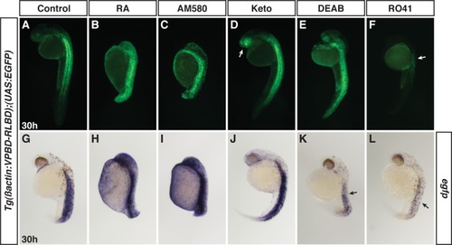
Tg(β-actin:VPBD-RLBD);(UAS:EGFP) embryos are sensitive to RAR agonists and antagonists. A, G: Control sibling embryos at 30 hpf. B, C, H, I: RA- and AM580-treated VP16-RA sensor embryos have almost ubiquitous expression. Embryos are posteriorized due to RAR agonist treatment. D, J: VP16-RA sensor embryos treated with Keto have expanded and enhanced expression, particularly in the anterior brain and eye (arrow in D). E, F, K, L: VP16-RA sensor embryos treated with DEAB or RO41. By ISH, DEAB and RO41 reduced expression (arrows in K and L). However, RO41 was more effective. By fluorescence, it was often hard to distinguish a loss in reporter expression in DEAB-treated embryos. While we observed reductions in reporter expression, particularly with RO41 treatment, we never completely lost expression in embryos treated with RAR or Aldh1a antagonists. All views are lateral with anterior up. |